. 2015 Jul;220(4):2449-68.
doi: 10.1007/s00429-014-0800-2. Epub 2014 Jun 3.

Reorganization of supramammillary-hippocampal pathways in the rat pilocarpine model of temporal lobe epilepsy: evidence for axon terminal sprouting

Affiliations

Reorganization of supramammillary-hippocampal pathways in the rat pilocarpine model of temporal lobe epilepsy: evidence for axon terminal sprouting

Rabia Soussi et al. Brain Struct Funct. 2015 Jul.

Abstract

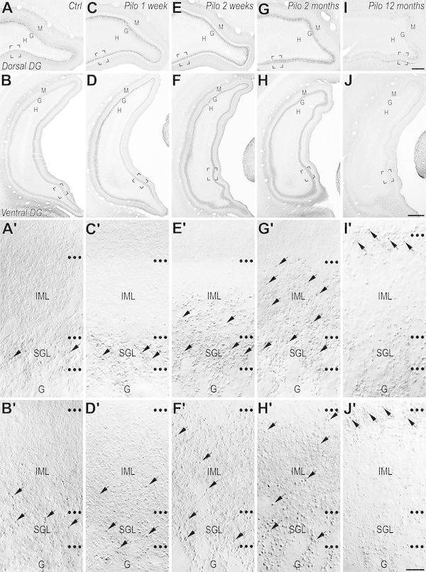
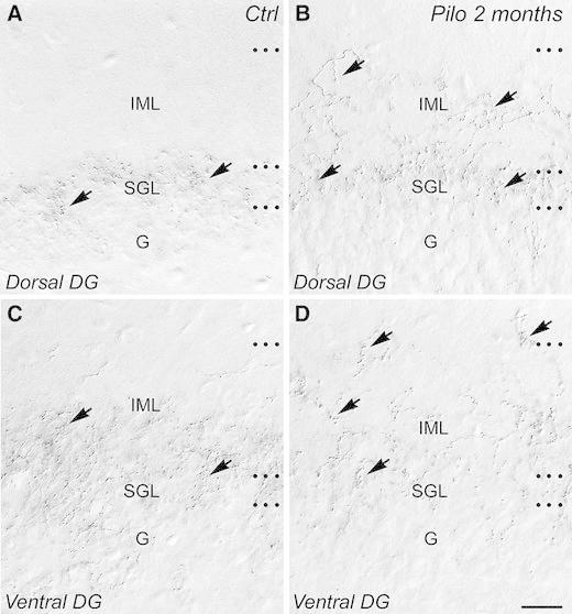
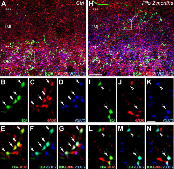
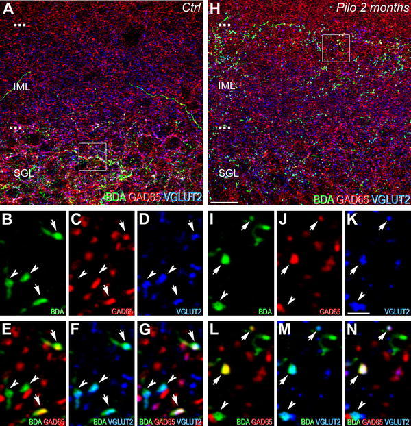
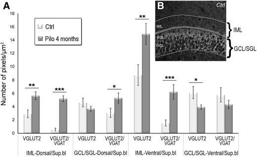
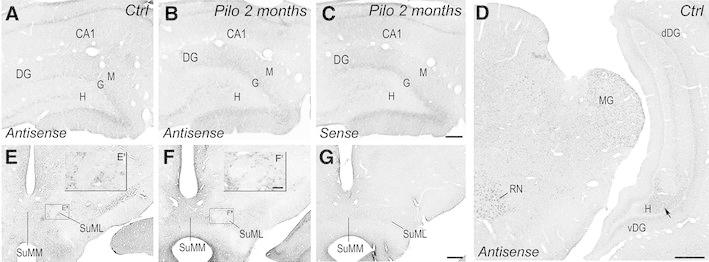
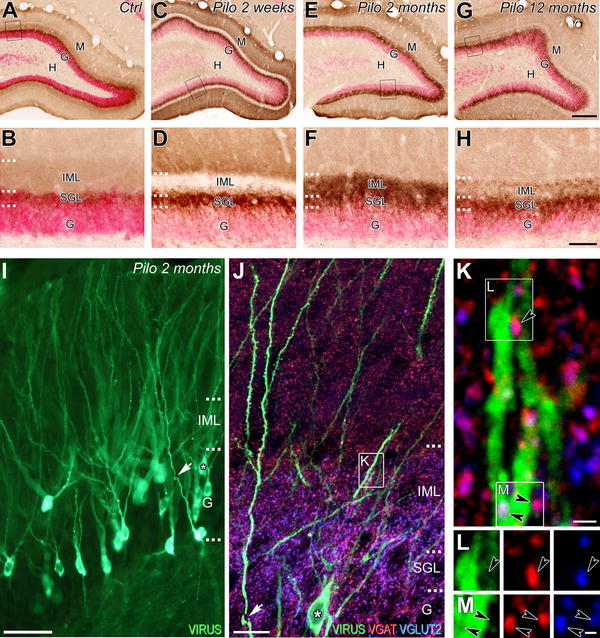
In mesial temporal lobe epilepsy (MTLE), spontaneous seizures likely originate from a multi-structural epileptogenic zone, including several regions of the limbic system connected to the hippocampal formation. In this study, we investigate the structural connectivity between the supramammillary nucleus (SuM) and the dentate gyrus (DG) in the model of MTLE induced by pilocarpine in the rat. This hypothalamic nucleus, which provides major extracortical projections to the hippocampal formation, plays a key role in the regulation of several hippocampus-dependent activities, including theta rhythms, memory function and emotional behavior, such as stress and anxiety, functions that are known to be altered in MTLE. Our findings demonstrate a marked reorganization of DG afferents originating from the SuM in pilocarpine-treated rats. This reorganization, which starts during the latent period, is massive when animals become epileptic and continue to evolve during epilepsy. It is characterized by an aberrant distribution and an increased number of axon terminals from neurons of both lateral and medial regions of the SuM, invading the entire inner molecular layer of the DG. This reorganization, which reflects an axon terminal sprouting from SuM neurons, could contribute to trigger spontaneous seizures within an altered hippocampal intrinsic circuitry.


Figures

Fig. 1
Fig. 1
Comparison of immunohistochemical labeling for VGLUT2 in coronal sections through the rostro-caudal extent of the dentate gyrus from control (a, b′) and pilocarpine-treated animals at 1 week (c, d′), 2 weeks (e, f′), 2 months (g, h′) and 12 months (i, j′). a′–j′ panels correspond to high magnification of the region outline in panels aj, respectively. ab′ In a control rat, VGLUT2 immunolabeling was present in the granular (G) and molecular (M) layers of the dentate gyrus (DG) at rostral (a) and caudal (b) levels. Virtually no labeling was observed in the hilus (H). At high magnification (a′, b′), VGLUT2 labeling included both punctate structures (representative examples pointed by arrows) and a diffuse labeling. The punctate structures, presumed axon terminals from the supramammillary nucleus neurons, displayed different patterns of distribution along the dorsal to ventral axis of the dentate gyrus (a, b). These terminals were concentrated in the supragranular layer (SGL) of the dorsal region of DG (a′) and were much sparser distributed throughout the inner molecular layer (IML) in the ventral DG (b′). The diffuse immunolabeling for VGLUT2 was observed in the inner and outer one-third of the molecular layer (a, b). cc′ In a pilocarpine-treated rat at 1 week, a decrease of the diffuse immunolabeling for VGLUT2 was evident in the IML of the dorsal (c, c′) but not in the ventral (d, d′) DG. As in control (a′, b′), many VGLUT2-containing terminals (arrows) were present in the SGL of the dorsal (c′) and ventral DG (d′). ef′ In a pilocarpine-treated rat at 2 weeks, the loss of VGLUT2 diffuse labeling was still evident in the IML of the dorsal DG (e, e′). In addition to the numerous VGLUT2-containing terminals (arrows) observed in the SGL, many of them were also present in the IML in the dorsal DG (arrows; e′) and ventral IML (arrows; f′). gh′ In an epileptic pilocarpine-treated rat at 2 months, numerous VGLUT2-containing terminals (arrows) were present in the entire IML throughout the rostro-caudal (g, h′) extent of the DG. An apparent recovery of diffuse labeling was observed in IML of the dorsal DG (g′). ij′ In an epileptic animal at 12 months, VGLUT2 immunolabeling was clearly different from that observed in control and pilocarpine-treated rats at 1 and 2 weeks but also from epileptic animals at 2 months. VGLUT2-containing terminals displayed a double-band distribution pattern, these terminals being located in the SGL and in the uppermost part of the IML (arrows). A marked loss of the diffuse immunolabeling for VGLUT2 was observed now throughout the entire rostro-caudal level in the IML including in the ventral dentate gyrus. H hilus, G granule cell layer, M molecular layer, IML inner molecular layer, Ctrl control, Pilo 1 week pilocarpine-treated animal at 1 week after SE, Pilo 2 weeks pilocarpine-treated animal at 2 weeks after SE, Pilo 2 months pilocarpine-treated animal at 2 months after SE, Pilo 12 months pilocarpine-treated animal at 12 months after SE. Scale bars 200 µm in a, c, e, g, i; 500 µm in b, d, f, h, j and 10 µm in a′–j
Fig. 2
Fig. 2
Comparison of the supramammillary–dentate gyrus pathways in coronal sections of dorsal (a, b) and ventral (c, d) dentate gyrus from control (a, c) and epileptic pilocarpine-treated animals (b, d) revealed with BDA anterograde tracing. ad In control as in epileptic animals, the detection of BDA anterograde tracer was performed 10 days after its injection in a region including both the lateral and medial parts of the supramammillary nucleus (SuM). a, c In a control rat, BDA-containing fibers and axon terminals (arrows) were restricted to the supragranular layer (SGL) in dorsal DG (a), whereas they were more distributed within the IML in the ventral DG (c). b, d In an epileptic animal at 2 months after pilocarpine injection, an aberrant distribution of BDA-containing fibers and axon terminals was evident. In the dorsal DG (b), these fibers and axon terminals (arrows) were located not only in the SGL as in a control rat but also invaded the entire IML (compare with panel a). In the ventral DG (d), many BDA-containing fibers and axon terminals (arrows) were also observed in the uppermost part of the IML in contrast to control animal (compare with panel c). DG dentate gyrus, G granule cell layer, SGL supragranular layer, IML inner molecular layer, Ctrl control, Pilo 2 months pilocarpine-treated rat at 2 months after status epilepticus. Scale bar 20 µm
Fig. 3
Fig. 3
Comparison of neurochemical phenotypes for the dorsal dentate gyrus afferents from the SuM between control (ag) and epileptic pilocarpine-treated (hn) rats, characterized by simultaneous labeling for BDA anterograde tracer (green), GAD65 (red) and VGLUT2 (blue) in coronal sections. a Image corresponding to a maximum intensity projection of a confocal slice z-stack (22 optical slices, spaced at 285 nm) showing labeling for BDA (green), GAD65 (red) and VGLUT2 (blue) in the dorsal DG of a control rat. Axon terminals and fibers, originating from SuM neurons and labeled for the BDA anterograde tracer (green), were located mainly in the SGL. Numerous GAD65-containing terminals (red) were present in the IML and SGL. VGLUT2-containing terminals (blue) were mainly located in the SGL. bd Images of the three different fluorophores used for the triple labeling, obtained by sequential acquisition of separate wavelength channels from a single confocal slice in the SGL of the dorsal DG demonstrated that many if not all axon terminals labeled for BDA (b, green, arrows) contained GAD65 (c, red, arrows) and VGLUT2 (d, blue, arrows). e Merge of b and c. f Merge of b and d. g Merge of bd. Triple-labeled boutons for BDA, GAD65 and VGLUT2 (white, arrows) were surrounded by double-labeled terminals for GAD65 and VGLUT2 (purple) as well as single-labeled terminals for GAD65 (red) or VGLUT2 (blue). h Image corresponding to a maximum intensity projection of a confocal slice z-stack (22 optical slices, spaced at 285 nm) showing labeling for BDA (green), GAD65 (red) and VGLUT2 (blue) in the dorsal DG of an epileptic rat at 2 months after pilocarpine injection. Axon terminals and fibers, originating from SuM neurons, labeled for the BDA anterograde tracer (green) were distributed within the entire IML in contrast to the control rat (compare with panel a). Numerous GAD65-containing terminals (red) were present in the IML and SGL. VGLUT2-containing terminals (blue) were located in the SGL but also in all the IML. ik Images of the three different fluorophores used for the triple labeling, obtained by sequential acquisition of separate wavelength channels from a single confocal slice, in the IML of the dorsal DG demonstrated two types of BDA-labeled axon terminals (i, green): the first one contained GAD65 (j, red, arrow) and VGLUT2 (k, blue, arrow), the second one contained VGLUT2 only (ik, arrowhead). l Merge of i and j. m Merge of i and k. n Merge of ik. Triple-labeled boutons for BDA, GAD65 and VGLUT2 (white, arrow) were surrounded by double-labeled terminals for BDA and VGLUT2 (arrowhead) as well as single-labeled terminals for GAD65 (red) or VGLUT2 (blue). Ctrl control, Pilo 2 months pilocarpine-treated rat at 2 months after status epilepticus. Scale bars 10 µm in a and h; 2 µm in bg and in
Fig. 4
Fig. 4
Comparison of neurochemical phenotypes for the ventral dentate gyrus afferents from the SuM between control (ag) and epileptic pilocarpine-treated (hn) rats, characterized by simultaneous labeling for BDA anterograde tracer (green), GAD65 (red) and VGLUT2 (blue) in coronal sections. a Image corresponding to a maximum intensity projection of a confocal slice z-stack (40 optical slices, spaced at 300 nm) showing labeling for BDA (green), GAD65 (red) and VGLUT2 (blue) in the ventral DG of a control rat. Axon terminals and fibers, originating from the SuM neurons, labeled for the BDA anterograde tracer (green) were distributed in the SGL and in the lower part of the IML. Numerous GAD65-containing terminals and VGLUT2-containing terminals were present in this region. bd Images of the three different fluorophores used for the triple labeling, obtained by sequential acquisition of separate wavelength channels from a single confocal slice, in the SGL of the ventral DG demonstrated axon terminals labeled for BDA (b, green, arrows) containing GAD65 (c, red, arrows) and VGLUT2 (d, blue, arrows) and BDA-labeled terminals containing VGLUT2 only (d, blue, arrowheads). e Merge of b and c. f Merge of b and d. g Merge of bd. Triple-labeled boutons for BDA, GAD65 and VGLUT2 (white, arrows) and double-labeled boutons for BDA and VGLUT2 (arrowheads) were surrounded by double-labeled terminals for GAD65 and VGLUT2 (purple) as well as single-labeled terminals for GAD65 (red) or VGLUT2 (blue). h Image corresponding to a maximum intensity projection of a confocal slice z-stack (40 optical slices, spaced at 300 nm) showing labeling for BDA (green), GAD65 (red) and VGLUT2 (blue) in the ventral DG of an epileptic rat at 2 months after pilocarpine injection. Axon terminals and fibers, originating from SuM neurons, labeled for the BDA anterograde tracer (green) displayed an aberrant distribution as compared to the control rat (a), many boutons and fibers being observed in the entire IML including the upper part. Numerous GAD65-containing terminals (red) and VGLUT2-containing terminals (blue) were also present in this entire region. ik Images of the three different fluorophores used for the triple labeling, obtained by sequential acquisition of separate wavelength channels from a single confocal slice, in the IML of the ventral DG demonstrated that many of these ectopic BDA-labeled axon terminals (i, green, arrows) contained GAD65 (j, red, arrows) and VGLUT2 (k, blue, arrows) and some contained VGLUT2 only (ik arrowhead). l Merge of i and j. m Merge of i and k. n Merge of ik. Triple-labeled boutons for BDA, GAD65 and VGLUT2 (white, arrows) and double-labeled terminals for BDA and VGLUT2 (arrowhead) were surrounded by double-labeled terminals for GAD65 and VGLUT2 (purple) as well as single-labeled terminals for GAD65 (red). Ctrl control, Pilo 2 months pilocarpine-treated rat at 2 months after status epilepticus. Scale bars 10 µm in a and h; 2 µm in bg and in
Fig. 5
Fig. 5
Quantitative analysis of VGLUT2 and VGAT proteins. a, b Quantitative analysis of the mean densities of labeling for VGLUT2 only and for VGLUT2/VGAT performed for the dorsal and ventral DG, in two regions of interest drawn over the inner molecular layer (IML) and granule cell layer which included the supragranular layer (GCL/SGL) as illustrated in (b) of the suprapyramidal blade (Sup.bl). Measures were obtained from three controls (white rectangles) and three pilocarpine-treated rats at 4 months (gray rectangles). Statistically significant differences are indicated (*p < 0.05; **p < 0.01; ***p < 0.001; ANOVA test). Errors bars SEM
Fig. 6
Fig. 6
Comparison of labeling for VGLUT2 mRNA in coronal sections of hippocampal formation (ad) and of the SuM (eg) from control (a, d, e) and epileptic pilocarpine-treated rats (b, c, f, g), processed for the same color reaction time. Sections of the hippocampal formation from a control rat (a) and an epileptic rat at 2 months (b), processed with antisense RNA probe, showed a faint nonspecific labeling for VGLUT2 in the pyramidal and granule cell layers similar to that observed in a section from an epileptic rat processed for the control sense RNA probe (c). d A section of the hippocampal formation, at a caudal level in a control rat, showing that hilar neurons, presumed mossy cells, in the ventral DG (vDG) expressed low levels of VGLUT2 mRNA as compared to mesencephalic nuclei including the red nucleus (RN) and the medial geniculate (MG). Hilar neurons in the dorsal DG (dDG) did not display detectable level of VGLUT2 mRNA. e In a section from a control rat, processed with antisense RNA probe, VGLUT2 mRNA was expressed by many diencephalic neurons including neurons located in the medial part (SuMM) and lateral part (SuML) of the supramammillary nucleus (SuM) as illustrated at higher magnification in the inset (e′). f In a section from an epileptic rat at 2 months after pilocarpine injection, processed with antisense RNA probe, revealed similar pattern and intensity of labeling for VGLUT2 mRNA in the SuMM and SuML (see inset f′) as that observed in the control rat (compare with e, e′). g No labeling was observed in an adjacent section from an epileptic rat at 2 months after pilocarpine injection, processed with sense RNA probe. Ctrl control, Pilo 2 months pilocarpine-treated rat at 2 months after status epilepticus. Scale bars 100 µm in e f, g; 25 µm in e′, f′; 50 µm in ac; 500 µm in d
Fig. 7
Fig. 7
Synaptic targets of ectopic dentate gyrus afferents from SuM neurons in pilocarpine-treated rats. ah Comparison of distribution patterns of VGLUT2-containing terminals (brown) and NeuN-labeled dentate granule cells (red) in coronal sections of the dorsal DG from a control rat (a, b) and pilocarpine-treated animals at 2 weeks (c, d), 2 months (e, f) and 12 months (g, h). b, d, f, h High magnifications of outlined areas, respectively, illustrated in panels a, c, e and g. In pilocarpine-treated rats (ch), the distribution pattern of dentate granule cell somata was similar to that observed in the control animal (a, b). No dispersion or bi-lamination of granule cells was associated with the aberrant VGLUT2-immunolabeling observed in the IML in pilocarpine-treated rat from 2 weeks on. i A section of dorsal DG from an epileptic animal at 2 months that was injected into the CA3 stratum lucidum of the dorsal hippocampus with the Rabies virus (RV) retrograde tracer to label cell bodies and dendritic trees of dentate granule cells (G). jk Image corresponding to a maximum intensity projection of a confocal slice z-stack (20 optical slices, spaced at 296 nm) showing labeling for the RV (green), VGAT (red) and VGLUT2 (blue) in the dorsal dentate gyrus of an epileptic rat at 2 month after pilocarpine injection. j High magnification of the region indicated by an arrow and a star in panel (i). Presumed axon terminals from SuM neurons, labeled for VGAT and VGLUT2, displayed an aberrant distribution with many boutons present in the entire IML including the upper part. Note that in the epileptic rat, the proximal apical dendrites (arrow) of dentate granule cells (*) across the IML displayed a low number of spines as compared to more distal segment. k Higher magnification of the outlined region indicated in panel j showing a dendritic tree of dentate granule cell across the upper IML contacted by ectopic VGLUT2/VGAT-containing axon terminals. l, m Images of the three different fluorophores used for the triple labeling, obtained by sequential acquisitions of separate wavelength channels from a single confocal slice, in the two outlined regions of the IML indicated in panel k and demonstrating that ectopic axon terminals labeled for VGAT (red, arrowhead) and VGLUT2 (blue, arrowhead), presumably originating from the SuM establish contact on the dendritic shafts of dentate granule cells retrogradely labeled with rabies virus (green). Scale bars 200 µm in a, c, e and g; 50 µm in b, d, f and h; 100 µm in i; 15 µm in j; 1 µm in k; 0.5 µm in l, m

References

    1. Abràmoff MD, Magelhaes PJ, Ram SJ. Image processing with ImageJ. Biophotonics Int. 2004;11:36–42.
    1. Barroso-Chinea P, Castle M, Aymerich MS, Perez-Manso M, Erro E, Tunon T, Lanciego JL. Expression of the mRNAs encoding for the vesicular glutamate transporters 1 and 2 in the rat thalamus. J Comp Neurol. 2007;501:703–715. doi: 10.1002/cne.21265. - DOI - PubMed
    1. Bettus G, Wendling F, Guye M, Valton L, Regis J, Chauvel P, Bartolomei F. Enhanced EEG functional connectivity in mesial temporal lobe epilepsy. Epilepsy Res. 2008;81:58–68. doi: 10.1016/j.eplepsyres.2008.04.020. - DOI - PubMed
    1. Blümcke I, Suter B, Behle K, Kuhn R, Schramm J, Elger CE, Wiestler OD. Loss of hilar mossy cells in Ammon’s horn sclerosis. Epilepsia. 2000;41:174–180. doi: 10.1111/j.1528-1157.2000.tb01577.x. - DOI - PubMed
    1. Boulland JL, Ferhat L, Tallak Solbu T, Ferrand N, Chaudhry FA, Storm-Mathisen J, Esclapez M. Changes in vesicular transporters for gamma-aminobutyric acid and glutamate reveal vulnerability and reorganization of hippocampal neurons following pilocarpine-induced seizures. J Comp Neurol. 2007;503:466–485. doi: 10.1002/cne.21384. - DOI - PubMed

Publication types

MeSH terms

LinkOut - more resources