. 2014 May:456-457:1-19.
doi: 10.1016/j.virol.2014.03.002. Epub 2014 Mar 25.

Endocytosis-mediated HIV-1 entry and its significance in the elusive behavior of the virus in astrocytes

Affiliations

Endocytosis-mediated HIV-1 entry and its significance in the elusive behavior of the virus in astrocytes

Ashok Chauhan et al. Virology. 2014 May.

Abstract

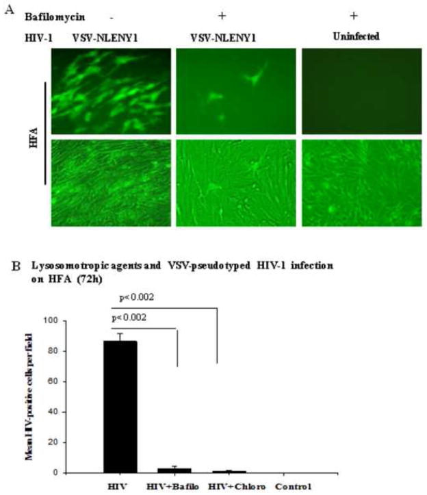
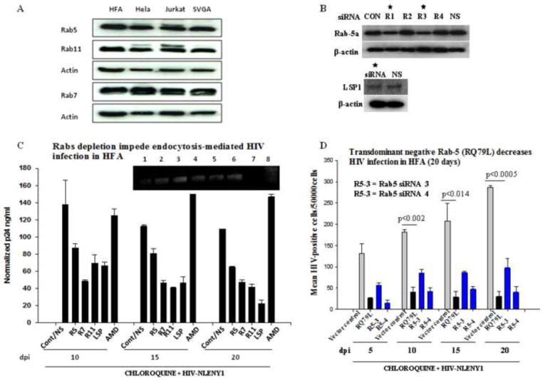
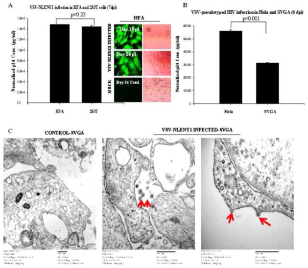
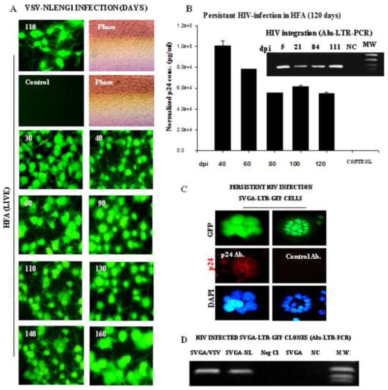
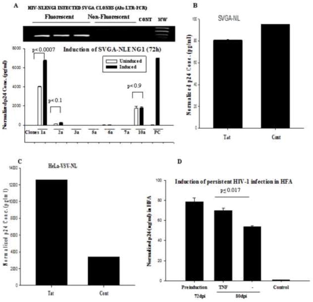
Astrocytes protect neurons but also evoke a proinflammatory response to injury and viral infections including HIV. We investigated the mechanism of HIV-1 infection in primary astrocytes, which showed minimal but productive viral infection independent of CXCR4. As with ectopic-CD4-expressing astrocytes, lysosomotropic agents led to increased HIV-1 infection in wild-type but not Rabs 5, 7, and 11-ablated astrocytes. Instead, HIV-1 infection was decreased in Rab-depleted astrocytes, corroborating viral entry by endocytosis. HIV-1 produced persistent infection in astrocytes (160 days); no evidence of latent infection was seen. Notably, one caveat is that endosomal modifiers enhanced wild-type HIV-1 infection (M- and T-tropic) in astrocytes, suggesting endocytic entry of the virus. Impeding endocytosis by inhibition of Rab 5, 7 or 11 will inhibit HIV infection in astrocytes. Although the contribution of such low-level infection in astrocytes to neurological complications is unclear, it may serve as an elusive viral reservoir in the central nervous system.

Keywords: HIV-1 persistence; HIV-LTR; LSP1; Lysosomotropic agents; Proteasome-inhibitors; Rab; TNPO3.


Figures

Figure 1
Figure 1. HIV-1 infection in astrocytes
(A) HIV-1 infection in Jurkat (NLENY1), primary macrophages (Ba-L), and primary human fetal (HFA) astrocytes (NLENY1). Cells were infected with HIV-1, then the culture supernatants from infected or uninfected cells were collected, and analyzed for viral p24 (ELISA). p24 levels in ng/ml, Jurkat (7dpi), macrophages (12 dpi), and HFA (15 dpi) are shown. (B) Diagrammatic view of HIV-1 infectious molecular clones, NL4-3 (wild-type), genetically engineered NL4-3 showing YFP (NLENY1) insertion and NLYUV3 (R-tropic) wherein V3 region of NL4-3 is replaced with V3 of HIV-1 YU-2. (C) HIV-1 infection in HFA: Upper panel shows productive NLENY1 infection after 7 days in Jurkat cells (positive control). Top second panel: HFA infection with NLENY1 virus (1.0 μg/mL p24) showing YFP fluorescence at 10 days post infection (dpi). Top third panel: SVGA were infected with NLENY1 virus (1.0 μg/mL p24) showing infected (YFP positive) cells (5dpi). Bottom panel: SVGA cells transfected with NLENY1 infectious molecular clone or pEGFP vector were immunostained after 48 h for p24 protein. (D) NLENY1 infected HFA: upper panel shows HIV-1 infection (YFP-positive) in HFA (GFAP- positive); middle panel: NLENY1 infection in HFA showing YFP and p24 expression. Bottom panel: uninfected HFA as controls for p24 antibody and isotype IgG (negative control)
Figure 2
Figure 2. Ectopic CD4 expression in astrocytes increase HIV-1 infection
(A) HFA were transduced with either CD4-expressing or empty lentiviral particles, and 24 h later, were infected with NLENY1 virus. In parallel, mock-infected HFA cultures were used as control. Infected cultures were followed for 15 days. YFP-positive (HIV) HFA were counted in 10 low-power fields (lpf) and the mean plotted (p ≤ 0025). (B) YFP-positive (HIV-1 infected) cells in CD4 expressing and wild-type HFA were observed by UV microscope at 15 days after infection. (C) HFA were transfected with CD4-expression vector and 48 h later infected with NLENY1 (1.0 μg per mL p24). HIV-1 infected HFA or CD4-expressing HFA were monitored by counting YFP-positive cells upto Day 31 after infection in the entire dish in duplicate every 5th day and fold-increase was calculated. (D) HIV-1 infected CD4-expressing HFA secrete p24 in extracellular culture medium. HFA-transfected cells with empty or CD4-expressing vectors were infected with NLENY1 virus (1 μg/ml). Cultures were monitored up to 27 days to determine YFP expression by fluorescence and p24 in culture supernatants by ELISA.
Figure 3
Figure 3. NLENY1-virus-infected HFA transinfect lymphocytes
(A–B) HFA were transfected either with empty or CD4-expressing vectors and, 24 h later, infected with NLENY1 virus. After extensive washing at 15 days after infection, HFA were co-cultured with uninfected lymphocytic cells. (A–B) Cultures were observed for YFP-positive lymphocytic cells. NLENY1-infected HFA (19 or 33 days; A, B) showing transinfection to lymphocytic cells at (A) 4 and (B) 18 days after co-culture.
Figure 4
Figure 4. Endosomal modulators (lysosomotropic agents) regulate HIV-1 infection in astrocytes
(A–B) Two-month old HFA seeded overnight in 6-well plates were either pre- or post treated with chloroquine (2.5–5.0 μM) for 1 h, then infected for 2 h with either NLYUV3 (M-tropic) (A) or NLENY1(T-tropic) (B) with 1.0 μg/ml of p24 equivalent. Infection was monitored by YFP fluorescence in entire well in duplicate and plotted as fold-increase from untreated infected cultures. (C) HIV-1 infected HFA left untreated or treated with chloroquine were followed for 21 days. ELISA for viral p24 protein on culture supernatants from HIV-1 infected HFA was positive up to 10 days in both chloroquine (QC)-treated and untreated HIV-1-infected cultures. In first 10 days there was a pseudo-p24 peak that did not correlate with fluorescence microscopic data. (D) Untreated HFA or HFA treated with chloroquine or CXCR4 antibody (α-CXCR4), was infected with NLENY1 virus (1 μg/mL p24). Fifteen days later, supernatants were used to infect normal Jurkat cells. Eight days later, supernatants from infected Jurkat cells were collected and analyzed for p24 by ELISA. Uninfected control cultures were used to normalize p24 levels.
Figure 5
Figure 5. Lysosomotropic agents inhibit VSV-pseudotyped HIV-1 infection in HFA
(A) HFA seeded overnight in six-well culture plates were infected with VSV-NLENY1 virus for 2 h, then washed two times with medium. Infected or uninfected cultures were left untreated or treated either with chloroquine (10 μM) or bafilomycin A (100 nM). Cultures were observed for 7 days for fluorescence; YFP-positive cells were observed by UV microscope and photographed. (B) HIV-positive fluorescent (YFP+) cells from (A) were counted in 7–9 fields in duplicate and plotted as mean ± SEM (p ≤ 0.002).
Figure 6
Figure 6. Depletion of Rab proteins in astrocytes obliterates endocytosis-mediated HIV-1 infection
(A) Astrocytes (HFA and SVGA) and HIV-permissive cells (lymphocytic and HeLa cells) grown for 48 h were processed for Western blot using monoclonal antibodies against Rab-5, -7 and -11. (B) Screenings of several siRNAs for depletion of each Rab and LSP1 (50–200 nM) were investigated on HFA by Western blotting. HFA were transfected with 200 nM each siRNA and, 72 h later lysates were analyzed by Western blotting for Rabs. Beta actin was used as a loading control. LSP1 siRNA was tested as a positive control to inhibit HIV-1 and non specific (NS) siRNA as a negative control. (C) Seeded HFA were transfected with 200 nM Rab specific siRNAs in parallel with LSP-siRNA and non-specific siRNA as controls. Then, one set of HFA was treated with CXCR4 blocker AMD3100 (AMD) before HIV-1 infection. At 72 h after siRNA transfection, HFA were infected with NLENY1 virus for 2 h. After washing, all cultures were treated with chloroquine (10 μM). At 48 h after infection (5 days after initial siRNA transfection), infected HFA were re-transfected with the respective siRNAs. p24 levels in culture supernatants collected at 10, 15, and 20 days after infection were analyzed by ELISA (n=4). HFA at 20 days after infection were tested for viral DNA integration by Alu-HIV LTR PCR (inset C). (D) HFA in 6-well culture plates were transfected with control vector, Rab 5a transdominant negative vectors, or Rab 5a siRNA #3 and #4 and, 72 h later infected with NLENY1. After washing, cultures were treated with chloroquine. Cultures were followed for 20 days after infection. YFP-positive HFA were counted in the entire well in duplicate and plotted (n=2).
Figure 7
Figure 7. Conducive intracellular environment for HIV-1 replication in astrocytes
(A) Late HIV-1 gene expression in HFA and SVGA upon transfection with HIV-1 infectious molecular clone. SVGA, SVGA-LTR-RFP reporter cells, or HFA were transfected with molecular HIV-1 clone (YU 2). In controls, empty vector DNA was transfected. At 48 h after transfection, cells were immunostained with monoclonal antibody for Nef (green) and observed under a UV microscope using a double filter (green/red). Left top panel: Nef immunostaining in SVGA-reporter cells; Left second panel: Nef expression in SVGA cells. Left third panel: Nef expression in HFA. Left fourth and bottom panels: HIV-1 late gene (p24) expression in SVGA and HFA. Cultured SVGA and HFA in flaskets were transfected with HIV-1 infectious molecular clone (YU-2) and, 48 h later, immunostained for p24 using monoclonal antibody (NIH). Panels on the right show p24 antibody on normal astrocytes as control. (B) Cultured SVGA and HeLa cells in six-well plates were transfected with 1 μg HIV-1 molecular clone of NL4-3, mutant NL4-3 (Δtat), or empty control vector; 72 h later, cell lysates were prepared. After protein normalization, Western blot analysis for viral Nef and p24 proteins was done in parallel with analysis for beta actin on the same blots.
Figure 8
Figure 8. Robust HIV-1 late gene expression in astrocytes
(A) HFA cultured for 48 h were infected with VSV-pseudotyped NL4-3 virus (200 ng/ mL p24) and, 48 h after infection, were immunostained for GFAP, p24 and nuclei. Images were captured under a fluorescence microscope (inset). Cells positive for both GFAP (red) and p24 (green) were randomly counted in 10 fields with total nuclear counts (blue) in duplicate sets; mean ± SEM were plotted using a sigma plot. (B) VSV-pseudotyped HIV-1 infection in SVGA-LTR reporter cells: Stable SVGA-LTR GFP or LTR-gagGFP-RRE reporter cells were seeded overnight in six-well culture plates. Next day, cells were infected with VSV-NLER+ virus for 2 h (200 ng/ mL p24) and washed. At 48 h after infection, cells were immunostained for p24 (red) and nuclei (blue). Dual-positive cells (green and red) were counted in ten random fields in duplicate cultures and mean positive cells ± SEM were plotted.
Figure 9
Figure 9. Productive HIV-1 infection in primary astrocytes
(A) HFA and 293T cells were infected with VSV-pseudotyped NLENY1 virus (100 ng/ mL p24). In parallel, untreated HFA and 293T cells were used as mock controls for normalization of p24 levels in the supernatants. Infected and uninfected culture supernatants from HFA and 293T cells at 7 days after infection were monitored for p24 by ELISA (p ≤ 0.23). Infected HFA were observed under UV microscope (inset). (B) Hela and SVGA cells were infected with VSV-pseudotyped-NL4-3 (100 ng/mL p24). Six days later, supernatants from infected and uninfected cells were monitored for p24 by ELISA (p ≤ 0.001). (C) HFA were infected with VSV-pseudotyped HIV-1 and, 48 h later, processed for transmission electron microscopy (TEM). Mock-infected HFA were used as control. Virus particles of 150–250 nm were observed intracellularly or at the membranes (40,000 magnifications).
Figure 10
Figure 10. Persistent HIV-1 infection in astrocytes
HFA in T-25 flasks were infected with VSV-NLENY1 virus and followed for 160 days. HIV-1 infection was monitored by YFP expression in infected HFA and p24 secretion in culture supernatants (A, B). (A) VSV-NLENY1- infected HFA showing YFP expression (live). (B) Viral kinetics in long-term (40 to 120 days) HIV-1 infected HFA is shown. Control (NC) is a mock-infected HFA. Alu-HIV-LTR PCR demonstrated HIV viral genome integration in infected HFA from 5 to 111 days after infection (inset B). (C) Persistent infection of HIV-1 in astrocytic (SVGA) cells: SVGA-LTR-GFP reporter cells were infected with HIV-1 NL4-3 and GFP-expressing cells were cloned. Clonal population of persistently HIV-1 (NLER+)-infected SVGA-LTR-GFP cells (chronic infection) showing sustained green fluorescence and p24 expression by immunofluorescence. (D) HIV-1 DNA integration by Alu-HIV-LTR PCR in NLER+ SVGA-LTRGFP infected cells is shown.
Figure 11
Figure 11. Reactivation of persistent HIV-1 infection in astrocytes
(A) Seven persistently HIV-1 infected clones of astrocytic cells (SVGA-NLENY1) were treated with TNF-α (10 ng/mL) for 72 h. In parallel, latently HIV-1 infected THP89-GFP cells were used a positive control. The supernatants were monitored for p24 by ELISA. One clone was inducible (p < 0.05); 6 were not (p ≤ 0.1, 0.9). (B–C) Persistently VSV-pseudotyped NLENY1-infected SVGA and Hela cells were cultured in six well plates and transfected with Tat or control vectors (0.5 μg per well). After 72 h, supernatants were analyzed for p24 in SVGA-NLENY1 (p ≤ 0.905) and Hela-NLENY1 cells (p ≤ 0.000005). (D) Persistently HIV-1-infected HFA were cultured overnight, then treated with TNF-α. At 48 h after treatment, supernatants from HFA were tested for p24 by ELISA. Controls were untreated HIV-1 infected HFA or TNF-a treated uninfected HFA.
Figure 12
Figure 12. Persistently HIV-1 infected astrocytes transinfect lymphocytes
(A) Persistently NLENY1-infected SVGA clone-1a or clone-10a was co-cultured with Jurkat cells for 7 days (cell-free wild-type HIV-1 normally infects Jurkat cells between 3–7 days). In controls, SVGA-pcDNA (stable) cells were co-cultured with uninfected lymphocytic cells; persistently infected HIV-1-infected cells served as base control. (B) The supernatants from (A) were analyzed for p24 by ELISA (p < 0.001, p < 0.0009).

Similar articles

Cited by

References

    1. Adachi A, Gendelman HE, Koenig S, Folks T, Willey R, Rabson A, Martin MA. Production of acquired immunodeficiency syndrome-associated retrovirus in human and nonhuman cells transfected with an infectious molecular clone. J Virol. 1986;59:284–291. - PMC - PubMed
    1. Beausejour Y, Tremblay MJ. Interaction between the cytoplasmic domain of ICAM-1 and Pr55Gag leads to acquisition of host ICAM-1 by human immunodeficiency virus type 1. J Virol. 2004;78:11916–11925. - PMC - PubMed
    1. Bencheikh M, Bentsman G, Sarkissian N, Canki M, Volsky DJ. Replication of different clones of human immunodeficiency virus type 1 in primary fetal human astrocytes: enhancement of viral gene expression by Nef. J Neurovirol. 1999;5:115–124. - PubMed
    1. Bounou S, Giguere JF, Cantin R, Gilbert C, Imbeault M, Martin G, Tremblay MJ. The importance of virus-associated host ICAM-1 in human immunodeficiency virus type 1 dissemination depends on the cellular context. Faseb J. 2004;18:1294–1296. - PubMed
    1. Boutet A, Salim H, Taoufik Y, Lledo PM, Vincent JD, Delfraissy JF, Tardieu M. Isolated human astrocytes are not susceptible to infection by M- and T-tropic HIV-1 strains despite functional expression of the chemokine receptors CCR5 and CXCR4. Glia. 2001;34:165–177. - PubMed

Publication types

Substances