. 2014 May:456-457:96-107.
doi: 10.1016/j.virol.2014.03.017. Epub 2014 Apr 3.

Abrogation of TLR3 inhibition by discrete amino acid changes in the C-terminal half of the West Nile virus NS1 protein

Affiliations

Abrogation of TLR3 inhibition by discrete amino acid changes in the C-terminal half of the West Nile virus NS1 protein

Clayton R Morrison et al. Virology. 2014 May.

Abstract

West Nile virus (WNV) is a mosquito-transmitted pathogen, which causes significant disease in humans. The innate immune system is a first-line defense against invading microorganism and many flaviviruses, including WNV, have evolved multifunctional proteins, which actively suppress its activation and antiviral actions. The WNV non-structural protein 1 (NS1) inhibits signal transduction originating from Toll-like receptor 3 (TLR3) and also critically contributes to virus genome replication. In this study we developed a novel FACS-based screen to attempt to separate these two functions. The individual amino acid changes P320S and M333V in NS1 restored TLR3 signaling in virus-infected HeLa cells. However, virus replication was also attenuated, suggesting that the two functions are not easily separated and may be contained within overlapping domains. The residues we identified are completely conserved among several mosquito- and tick-borne flaviviruses, indicating that they are of biological importance to the virus.

Keywords: Flavivirus; Innate immunity; NS1; TLR3; West Nile virus.


Figures

Figure 1
Figure 1. Screen work-flow for the identification of mutagenized NS1 proteins that allow TLR3 signaling and support replication of a WNV NS1-deleted GFP replicon particle
A HeLa reporter cell library expressing random NS1 mutants (HeLa-NFκB-DsRed2-pLEX-mNS1) was infected at an MOI of 1 with NS1-deleted WNV GFP replicon particles (WNV ΔNS1 GFP VRP) for 24h followed by pIC treatment for an additional 36h. GFP and DsRed2 double-positive cells were sorted by Fluorescence-activated cell sorting (FACS), individual clones expanded and the NS1 sequence of each clone was determined after confirmation of the desired phenotype.
Figure 2
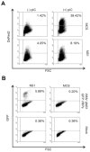
Figure 2. Validation of the tools employed by the screen
(A) HeLa-NFκB-DsRed2 cells stably transduced with either pLEX-NS1 or pLEX-MCS (vector control) lentivirus were treated with pIC for 36h or left untreated and DsRed2 expression was determined by flow cytometry. (B) HeLa-NFκB-DsRed2 cells stably transduced with either pLEX-NS1 or pLEX-MCS (vector control) lentivirus were either infected for 24h with WNV ΔNS1 GFP VRPs or mock infected. GFP expression was determined by flow cytometry. Data are representative of three independent experiments.
Figure 3
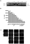
Figure 3. Efficiency of identified NS1 amino acid substitution proteins to trans-complement WNV ΔNS1 GFP VRPs
(A) HeLa-NFκB-luc reporter cells were transduced with lentivirus expressing either wt NS1, MCS, or single or double NS1 amino acid changes and selected for puromycin resistance. 5μg of whole cell lysate from each cell line was probed for NS1 expression by immunoblot and normalized to β-actin expression. (B) HeLa-NFκB-luc reporter cells stably transduced with lentivirus expressing either wt NS1, MCS, or single or double NS1 amino acid substitutions were infected at an MOI of 0.15 for 36h with WNV ΔNS1 GFP VRPs or mock infected. Expression of GFP was quantified by flow cytometry by determining the mean fluorescent intensity (MFI). The background MFI of identically infected MCS cells was subtracted from the MFI determined for other infected cells. Data are the average of two independent experiments and error bars represent standard deviation from the mean. (C) GFP expression due to trans-complementation by wild type and NS1 proteins with coding changes was monitored by fluorescence microscopy.
Figure 4
Figure 4. Efficiency of TLR3 signaling in cells expressing NS1 amino acid substitution proteins
HeLa-NFκB-luc reporter cells stably transduced with lentivirus expressing either wt NS1, MCS, or single or double NS1 amino acid changes were treated with 20μg/ml pIC for 8h or left untreated. (A) IL-6 secretion in the supernatants of cells was determined by enzyme-linked immunosorbent assay (ELISA) and fold induction was calculated by comparing IL-6 produced by pIC treated cells to that of untreated cells. Data are the average of two independent experiments performed in biological triplicate and error bars represent standard deviation from the mean. Statistical significance was determined by Mann-Whitney U test, where *p≤0.05 NS= not significant. All samples were compared to wt NS1. (B) Fold NFκB induction was determined by luciferase reporter assay. Relative light units were measured and reported as fold induction over untreated cells. Data are the average of two independent experiments and error bars represent standard deviation from the mean. Statistical significance was determined by student’s t-test, where *p≤0.05 NS= not significant. All samples were compared to wt NS1.
Figure 5
Figure 5. Shannon entropy (H) and multiple protein alignment of the NS1 proteins of several mosquito- and tick-borne flaviviruses
The amino acid sequences of 10 mosquito-borne and 6 tick-borne flaviviruses were aligned with ClustalW using the Blosum scoring matrix. The multiple-alignment file was displayed with BOXshade 3.21 where black-shading represents identical residues and gray-shading represents conserved substitutions. Shannon entropy analysis of the multiple protein alignment was performed by the Protein Variability Server (PVS) at the Complutense University of Madrid, and the data generated was displayed in MS Excel. H ≤ 1 are considered highly conserved residues. Mosquito-borne flaviviruses are represented in bolded font and tick-borne flaviviruses in italics.
Figure 6
Figure 6. Replication kinetics and TLR3 inhibition of mutant WNV expressing the identified amino acid residue changes in the NS1 protein
(A) HeLa cells were infected at an MOI of 0.01 with each WNV NS1 mutant and virus containing supernatants were collected every 24h for 96h. Infectious virus was titered on Vero cells by immunofocus assay. Growth curves were performed in biological triplicate and data are representative of two independent experiments. Error bars represent standard deviation from the mean. Statistical significance was determined by student’s t-test, where *p≤0.05. (B) HeLa cells were infected at an MOI of 5 with each WNV NS1 mutant for 1h, washed with PBS, replaced with fresh medium, and allowed to incubate for 20h. Medium was removed and replaced with medium with or without pIC (20μg/ml) for 8h. Cell culture supernatants were collected and IL-6 concentrations were determined by ELISA as pg of IL-6/ml. Data are representative of three independent experiments and error bars represent standard deviation from the mean. Statistical significance was determined by student’s t-test, where *p≤0.05 and NS= not significant. All samples were compared to mock infected cells treated with pIC. (C) HeLa cells infected with identical amounts of wt and mutant viruses were fixed and permeabilized 28h post-infection and infected cells were identified by flow cytometry using an E protein-specific primary monoclonal antibody and were detected with a secondary Alex Fluor 488 antibody. Grey filled histograms represent uninfected control cells and black open histograms represent E protein in WNV infected cell. Data are representative of three independent experiments.

Similar articles

Cited by

References

    1. Aguirre S, Maestre AM, Pagni S, Patel JR, Savage T, Gutman D, Maringer K, Bernal-Rubio D, Shabman RS, Simon V, Rodriguez-Madoz JR, Mulder LC, Barber GN, Fernandez-Sesma A. DENV inhibits type I IFN production in infected cells by cleaving human STING. PLoS Pathog. 2012;8(10):e1002934. - PMC - PubMed
    1. Alcon-LePoder S, Drouet MT, Roux P, Frenkiel MP, Arborio M, Durand-Schneider AM, Maurice M, Le Blanc I, Gruenberg J, Flamand M. The secreted form of dengue virus nonstructural protein NS1 is endocytosed by hepatocytes and accumulates in late endosomes: implications for viral infectivity. J Virol. 2005;79(17):11403–11. - PMC - PubMed
    1. Ashour J, Laurent-Rolle M, Shi PY, Garcia-Sastre A. NS5 of dengue virus mediates STAT2 binding and degradation. J Virol. 2009;83(11):5408–18. - PMC - PubMed
    1. Avirutnan P, Fuchs A, Hauhart RE, Somnuke P, Youn S, Diamond MS, Atkinson JP. Antagonism of the complement component C4 by flavivirus nonstructural protein NS1. J Exp Med. 2010;207(4):793–806. - PMC - PubMed
    1. Avirutnan P, Hauhart RE, Somnuke P, Blom AM, Diamond MS, Atkinson JP. Binding of flavivirus nonstructural protein NS1 to C4b binding protein modulates complement activation. J Immunol. 2011;187(1):424–33. - PMC - PubMed

Publication types

MeSH terms

Substances

LinkOut - more resources