Multicenter Study
. 2014 Jun 2:2:58.
doi: 10.1186/2051-5960-2-58.

A new sensitive PCR assay for one-step detection of 12 IDH1/2 mutations in glioma

Affiliations
Multicenter Study

A new sensitive PCR assay for one-step detection of 12 IDH1/2 mutations in glioma

Aurélie Catteau et al. Acta Neuropathol Commun. .

Abstract

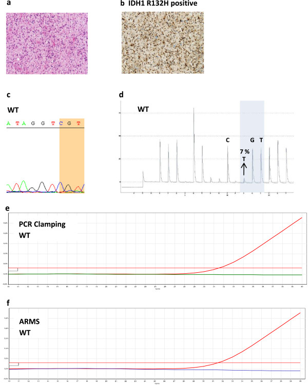
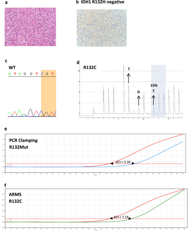
Introduction: Mutations in isocitrate dehydrogenase genes IDH1 or IDH2 are frequent in glioma, and IDH mutation status is a strong diagnostic and prognostic marker. Current IDH mutation screening is performed with an immunohistochemistry (IHC) assay specific for IDH1 R132H, the most common mutation. Sequencing is recommended as a second-step test for IHC-negative or -equivocal cases. We developed and validated a new real-time quantitative polymerase chain reaction (PCR) assay for single-step detection of IDH1 R132H and 11 rare IDH1/2 mutations in formalin-fixed paraffin-embedded (FFPE) glioma samples. Performance of the IDH1/2 PCR assay was compared to IHC and Sanger sequencing.

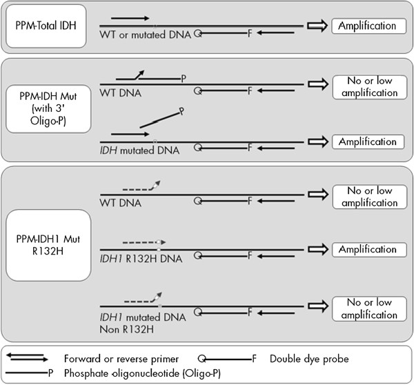
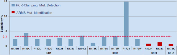
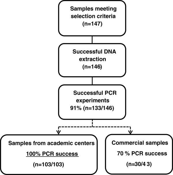
Results: The IDH1/2 PCR assay combines PCR clamping for detection of 7 IDH1 and 5 IDH2 mutations, and Amplification Refractory Mutation System technology for specific identification of the 3 most common mutations (IDH1 R132H, IDH1 R132C, IDH2 R172K). Analytical sensitivity of the PCR assay for mutation detection was <5% for 11/12 mutations (mean: 3.3%), and sensitivity for mutation identification was very high (0.8% for IDH1 R132H; 1.2% for IDH1 R132C; 0.6% for IDH2 R172K). Assay performance was further validated on 171 clinical glioma FFPE samples; of these, 147 samples met the selection criteria and 146 DNA samples were successfully extracted. IDH1/2 status was successfully obtained in 91% of cases. All but one positive IDH1 R132H-IHC cases were concordantly detected by PCR and 3 were not detected by sequencing. Among the IHC-negative cases (n = 72), PCR detected 12 additional rare mutations (10 IDH1, 2 IDH2). All mutations detected by sequencing (n = 67) were concordantly detected by PCR and 5/66 sequencing-negative cases were PCR-positive (overall concordance: 96%). Analysis of synthetic samples representative of the 11 rare IDH1/2 mutations detected by the assay produced 100% correct results.

Conclusions: The new IDH1/2 PCR assay has a high technical success rate and is more sensitive than Sanger sequencing. Positive concordance was 98% with IHC for IDH1 R132H detection and 100% with sequencing. The PCR assay can reliably be performed on FFPE samples and has a faster turnaround time than current IDH mutation detection algorithms. The assay should facilitate implementation of a comprehensive IDH1/2 testing protocol in routine clinical practice.


Figures

Figure 1
Figure 1
Principle of the IDH1/2 PCR assay. Total reaction mixes (top) The total Primers and Probe Mixes (PPM-Total) used primers and probes to amplify both mutated and wild-type (WT) target sequences. Mutation detection reaction mixes (middle) The mutation detection primers and probe mixes combined primers and probes, to amplify both mutated and WT target sequences, plus an oligonucleotide, 3'-blocked with the addition of a phosphate group (3’-Oligo-P) to prevent elongation (PCR clamping), which was specific to the WT target sequence. When the PCR template contained the WT sequence, the 3'-Oligo-P bound preferentially over the PCR primer binding due to higher affinity. There was no or low extension by the DNA polymerase and no or low amplification was observed. When a mutated sequence was present, PCR primer bound preferentially over the 3'-Oligo-P and amplification proceeded. Mutation identification reaction mixes (bottom) Allele-specific amplification was achieved by ARMS® (Amplification Refractory Mutation System), which exploits the ability of the DNA polymerase to distinguish between a match and a mismatch at the 3' end of a PCR primer. When the PCR primer fully matched, the amplification proceeded with full efficiency. When the 3' base was a mismatch, only low-level background amplification occurred. The same principle shown on the figure to detect IDH1 R132H applied for IDH1 R132C and IDH2 R172K.
Figure 2
Figure 2
Sensitivity of the IDH1/2 PCR assay for the 12 detected and 3 identified IDH1/2 mutations. The sensitivity defined as the lowest amount of mutant DNA in a background of wild-type DNA was indicated for each of the 12 IDH1/2 mutations detected with the PCR IDH1/2 kit. The cut-off values were derived from a total of 2250 ΔCt measurements. Sensitivity varied across mutations from 0.6% to 15% with a mean of 3.3% (red dotted line). Limit of Detection was <5% for 11/12 mutations and ≤3% for 9 of them.
Figure 3
Figure 3
Distribution of the clinical glioma samples by tumor type (World Health Organization grade) (n = 147). The majority of the collected formalin-fixed paraffin-embedded clinical samples meeting inclusion criteria for the IDH1/2 PCR test originated from patients with diagnosed primary glioblastoma, the most frequently diagnosed glioma subtype in clinical routine. The remaining samples originated from patients diagnosed with other histological forms, mainly astrocytomas. Samples were available for each diffuse glioma subtype.
Figure 4
Figure 4
IDH1/2 PCR assay experimental flowchart: technical success rate. Out of the 147 formalin-fixed paraffin-embedded glioma clinical samples meeting the selection criteria, one DNA extraction failed. From the remaining samples, IDH1/2 PCR assay results were successfully obtained for most of the samples (91%). Successful tests were achieved on all samples (n = 103) originating from academic centers.
Figure 5
Figure 5
Discordant case # 1. Case #1 stained IDH1 R132H positive by immunochemistry but was found WT for the IDH1 codon 132 (CGT) by Sanger sequencing, by pyrosequencing and with the IDH1/2 PCR assay a H&E slide. b IHC using the anti-human IDH1 R132H mouse monoclonal antibody DIA clone H09. c Sanger sequencing. d pyrogram indicating an allele frequency of 7%, thus below the cutoff for mutation ≥ 15%. e, f IDH1/2 PCR assay combining PCR-clamping (e) and ARMS (f): the red horizontal lines correspond to the threshold used to determine Ct. Total copies of IDH1 (WT ± mutated) are amplified with the total amplification reaction (red curves) but without further amplification by PCR-Clamping (blue curve) or by ARMS with specific R132H primers (green curve), indicating the absence of any mutation within IDH1 codon 132.
Figure 6
Figure 6
Discordant case #4. Case #4 was IDH1 R132H negative by IHC and was identified as wild-type by Sanger sequencing for IDH1 codon 132. The IDH1/2 PCR assay identified an IDH1 R132C mutation which was confirmed by pyrosequencing. a H&E slide. b IDH1 R132H IHC positive staining. c Sanger sequencing. d the pyrogram indicates the presence of the IDH1 R132C mutation (TGT) at a low allele frequency (around 15%) suggesting that the sample contained low tumor cell content. e PCR-clamping: the difference between the IDH1 PCR-clamping mutation assay Ct (blue curve) and total assay Ct (red curve) is inferior to the cutoff value (ΔCt = 31.87 - 27.49 = 4.38 < 5.34) indicating the presence of a mutation within IDH1 codon 132. f ARMS: the difference between the total assay Ct (red curve) and the IDH1 R132C ARMS assay Ct (green curve) is inferior to the cutoff value (ΔCt = 31.20 – 27.49 = 3.71 < 7.14) indicating presence of the IDH1 R132C mutation.

Similar articles

Cited by

References

    1. Louis DN, Ohgaki H, Wiestler OD, Cavenee WK, Burger PC, Jouvet A, Scheithauer BW, Kleihues P. The 2007 WHO classification of tumours of the central nervous system. Acta Neuropathol. 2007;114(2):97–109. doi: 10.1007/s00401-007-0243-4. - DOI - PMC - PubMed
    1. Weller M, Pfister SM, Wick W, Hegi ME, Reifenberger G, Stupp R. Molecular neuro-oncology in clinical practice: a new horizon. Lancet Oncol. 2013;14(9):e370–e379. doi: 10.1016/S1470-2045(13)70168-2. - DOI - PubMed
    1. Thomas L, Di Stefano AL, Ducray F. Predictive biomarkers in adult gliomas: the present and the future. Curr Opin Oncol. 2013;25(6):689–694. doi: 10.1097/CCO.0000000000000002. - DOI - PubMed
    1. Hartmann C, Meyer J, Balss J, Capper D, Mueller W, Christians A, Felsberg J, Wolter M, Mawrin C, Wick W, Weller M, Herold-Mende C, Unterberg A, Jeuken JW, Wesseling P, Reifenberger G, von Deimling A. Type and frequency of IDH1 and IDH2 mutations are related to astrocytic and oligodendroglial differentiation and age: a study of 1,010 diffuse gliomas. Acta Neuropathol. 2009;118(4):469–474. doi: 10.1007/s00401-009-0561-9. - DOI - PubMed
    1. von Deimling A, Korshunov A, Hartmann C. The next generation of glioma biomarkers: MGMT methylation, BRAF fusions and IDH1 mutations. Brain Pathol. 2011;21(1):74–87. doi: 10.1111/j.1750-3639.2010.00454.x. - DOI - PMC - PubMed

Publication types