

Processing multiple visual objects is limited by overlap in neural channels
- PMID: 24889618
- PMCID: PMC4066506
- DOI: 10.1073/pnas.1317860111
Processing multiple visual objects is limited by overlap in neural channels
Abstract
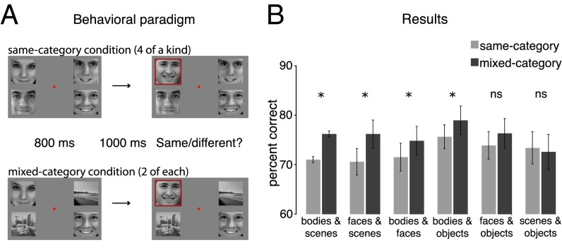
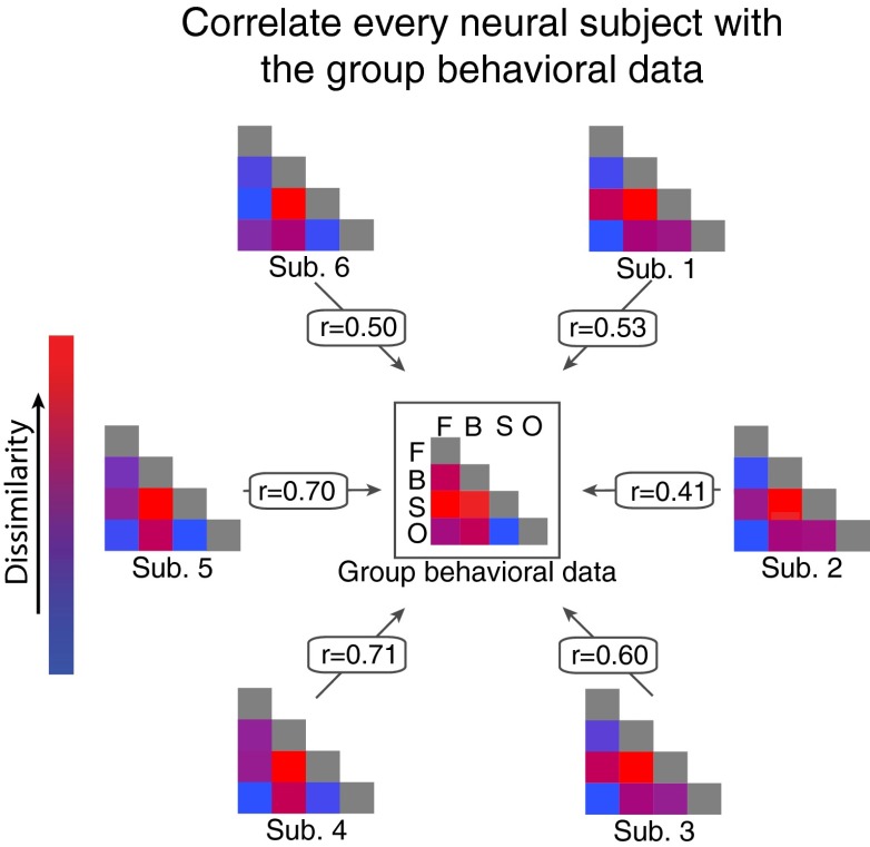
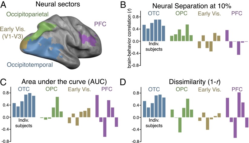
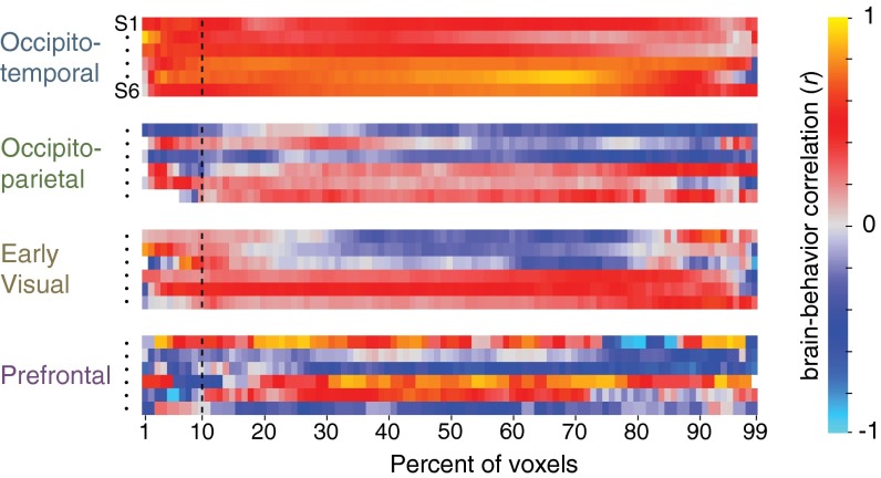
High-level visual categories (e.g., faces, bodies, scenes, and objects) have separable neural representations across the visual cortex. Here, we show that this division of neural resources affects the ability to simultaneously process multiple items. In a behavioral task, we found that performance was superior when items were drawn from different categories (e.g., two faces/two scenes) compared to when items were drawn from one category (e.g., four faces). The magnitude of this mixed-category benefit depended on which stimulus categories were paired together (e.g., faces and scenes showed a greater behavioral benefit than objects and scenes). Using functional neuroimaging (i.e., functional MRI), we showed that the size of the mixed-category benefit was predicted by the amount of separation between neural response patterns, particularly within occipitotemporal cortex. These results suggest that the ability to process multiple items at once is limited by the extent to which those items are represented by separate neural populations.
Keywords: capacity limitations; competition; representational similarity; visual cognition; working memory.
Conflict of interest statement
The authors declare no conflict of interest.
Figures

Similar articles
-
Visual object categorization in infancy.Proc Natl Acad Sci U S A. 2022 Feb 22;119(8):e2105866119. doi: 10.1073/pnas.2105866119. Proc Natl Acad Sci U S A. 2022. PMID: 35169072 Free PMC article.
-
Extrastriate cortex and medial temporal lobe regions respond differentially to visual feature overlap within preferred stimulus category.Neuropsychologia. 2012 Nov;50(13):3053-61. doi: 10.1016/j.neuropsychologia.2012.07.006. Epub 2012 Jul 20. Neuropsychologia. 2012. PMID: 22820343
-
A "Bandwidth" in cortical representations of multiple faces.Cereb Cortex. 2023 Sep 9;33(18):10028-10035. doi: 10.1093/cercor/bhad262. Cereb Cortex. 2023. PMID: 37522262
-
Visual Object Recognition: Do We (Finally) Know More Now Than We Did?Annu Rev Vis Sci. 2016 Oct 14;2:377-396. doi: 10.1146/annurev-vision-111815-114621. Epub 2016 Aug 3. Annu Rev Vis Sci. 2016. PMID: 28532357 Review.
-
Division of labor between lateral and ventral extrastriate representations of faces, bodies, and objects.J Cogn Neurosci. 2011 Dec;23(12):4122-37. doi: 10.1162/jocn_a_00091. Epub 2011 Jul 7. J Cogn Neurosci. 2011. PMID: 21736460 Review.
Cited by
-
Semantic Expectation Effects on Object Detection: Using Figure Assignment to Elucidate Mechanisms.Vision (Basel). 2022 Mar 21;6(1):19. doi: 10.3390/vision6010019. Vision (Basel). 2022. PMID: 35324604 Free PMC article.
-
Individual variation in the functional lateralization of human ventral temporal cortex: Local competition and long-range coupling.Imaging Neurosci (Camb). 2025 Mar 3;3:imag_a_00488. doi: 10.1162/imag_a_00488. eCollection 2025 Mar 1. Imaging Neurosci (Camb). 2025. PMID: 40078535 Free PMC article.
-
What is the Bandwidth of Perceptual Experience?Trends Cogn Sci. 2016 May;20(5):324-335. doi: 10.1016/j.tics.2016.03.006. Trends Cogn Sci. 2016. PMID: 27105668 Free PMC article. Review.
-
The Ventral Visual Pathway Represents Animal Appearance over Animacy, Unlike Human Behavior and Deep Neural Networks.J Neurosci. 2019 Aug 14;39(33):6513-6525. doi: 10.1523/JNEUROSCI.1714-18.2019. Epub 2019 Jun 13. J Neurosci. 2019. PMID: 31196934 Free PMC article.
-
The Effects of Similarity on High-Level Visual Working Memory Processing.Adv Cogn Psychol. 2017 Dec 31;13(4):296-305. doi: 10.5709/acp-0229-8. eCollection 2017. Adv Cogn Psychol. 2017. PMID: 29362645 Free PMC article.
References
-
- Baddeley AD, Hitch G. Working memory. In: Bower GH, editor. The Psychology of Learning and Motivation: Advances in Research and Theory. New York: Academic; 1974. pp. 47–89.
-
- Duncan J, Martens S, Ward R. Restricted attentional capacity within but not between sensory modalities. Nature. 1997;387(6635):808–810. - PubMed
-
- Gazzaniga M, Ivry RB, Mangun GR. Cognitive Neuroscience. New York: Norton; 2008.
Publication types
MeSH terms
Grants and funding
LinkOut - more resources
Full Text Sources
Other Literature Sources
