

Protons are a neurotransmitter that regulates synaptic plasticity in the lateral amygdala
- PMID: 24889629
- PMCID: PMC4066526
- DOI: 10.1073/pnas.1407018111
Protons are a neurotransmitter that regulates synaptic plasticity in the lateral amygdala
Abstract
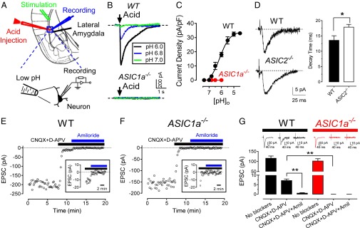
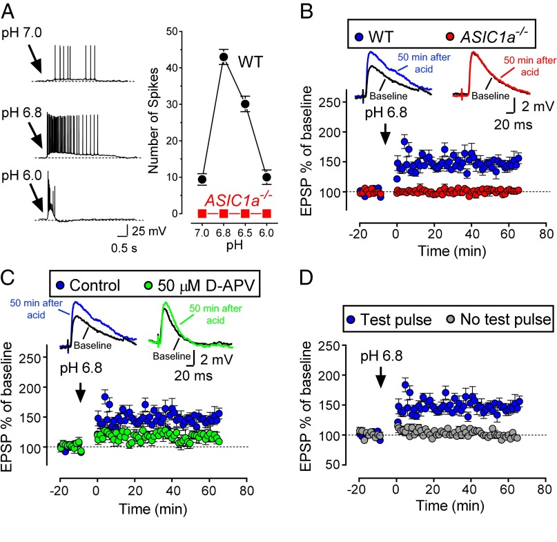
Stimulating presynaptic terminals can increase the proton concentration in synapses. Potential receptors for protons are acid-sensing ion channels (ASICs), Na(+)- and Ca(2+)-permeable channels that are activated by extracellular acidosis. Those observations suggest that protons might be a neurotransmitter. We found that presynaptic stimulation transiently reduced extracellular pH in the amygdala. The protons activated ASICs in lateral amygdala pyramidal neurons, generating excitatory postsynaptic currents. Moreover, both protons and ASICs were required for synaptic plasticity in lateral amygdala neurons. The results identify protons as a neurotransmitter, and they establish ASICs as the postsynaptic receptor. They also indicate that protons and ASICs are a neurotransmitter/receptor pair critical for amygdala-dependent learning and memory.
Keywords: PcTX1; acid sensing ion channel; long-term potentiation.
Conflict of interest statement
The authors declare no conflict of interest.
Figures
Similar articles
-
Acid-Sensing Ion Channels Activated by Evoked Released Protons Modulate Synaptic Transmission at the Mouse Calyx of Held Synapse.J Neurosci. 2017 Mar 8;37(10):2589-2599. doi: 10.1523/JNEUROSCI.2566-16.2017. Epub 2017 Feb 3. J Neurosci. 2017. PMID: 28159907 Free PMC article.
-
ASIC-dependent LTP at multiple glutamatergic synapses in amygdala network is required for fear memory.Sci Rep. 2015 May 19;5:10143. doi: 10.1038/srep10143. Sci Rep. 2015. PMID: 25988357 Free PMC article.
-
SK channels regulate excitatory synaptic transmission and plasticity in the lateral amygdala.Nat Neurosci. 2005 May;8(5):635-41. doi: 10.1038/nn1450. Epub 2005 Apr 24. Nat Neurosci. 2005. PMID: 15852010
-
Synaptic signals mediated by protons and acid-sensing ion channels.Synapse. 2019 Oct;73(10):e22120. doi: 10.1002/syn.22120. Epub 2019 Jul 15. Synapse. 2019. PMID: 31180161 Review.
-
Acid-Sensing Ion Channels Structural Aspects, Pathophysiological Importance and Experimental Mutational Data Available Across Various Species to Target Human ASIC1.Curr Drug Targets. 2019;20(1):111-121. doi: 10.2174/1389450119666180820103316. Curr Drug Targets. 2019. PMID: 30124148 Review.
Cited by
-
GPR68 Is a Neuroprotective Proton Receptor in Brain Ischemia.Stroke. 2020 Dec;51(12):3690-3700. doi: 10.1161/STROKEAHA.120.031479. Epub 2020 Oct 16. Stroke. 2020. PMID: 33059544 Free PMC article.
-
Two aspects of ASIC function: Synaptic plasticity and neuronal injury.Neuropharmacology. 2015 Jul;94:42-8. doi: 10.1016/j.neuropharm.2014.12.010. Epub 2015 Jan 9. Neuropharmacology. 2015. PMID: 25582290 Free PMC article. Review.
-
A Cytosolic Amphiphilic α-Helix Controls the Activity of the Bile Acid-sensitive Ion Channel (BASIC).J Biol Chem. 2016 Nov 18;291(47):24551-24565. doi: 10.1074/jbc.M116.756437. Epub 2016 Sep 27. J Biol Chem. 2016. PMID: 27679529 Free PMC article.
-
Peripheral and central employment of acid-sensing ion channels during early bilaterian evolution.Elife. 2023 Feb 23;12:e81613. doi: 10.7554/eLife.81613. Elife. 2023. PMID: 36821351 Free PMC article.
-
Neural substrates of habit.J Neurosci Res. 2020 Jun;98(6):986-997. doi: 10.1002/jnr.24552. Epub 2019 Nov 6. J Neurosci Res. 2020. PMID: 31693205 Free PMC article. Review.
References
-
- Füldner HH, Stadler H. 31P-NMR analysis of synaptic vesicles. Status of ATP and internal pH. Eur J Biochem. 1982;121(3):519–524. - PubMed
-
- Michaelson DM, Angel I. Determination of delta pH in cholinergic synaptic vesicles: Its effect on storage and release of acetylcholine. Life Sci. 1980;27(1):39–44. - PubMed
-
- Miesenböck G, De Angelis DA, Rothman JE. Visualizing secretion and synaptic transmission with pH-sensitive green fluorescent proteins. Nature. 1998;394(6689):192–195. - PubMed
-
- DeVries SH. Exocytosed protons feedback to suppress the Ca2+ current in mammalian cone photoreceptors. Neuron. 2001;32(6):1107–1117. - PubMed
Publication types
MeSH terms
Substances
Grants and funding
LinkOut - more resources
Full Text Sources
Other Literature Sources
Molecular Biology Databases
Miscellaneous
