

Apoptosis of alcohol-exposed human placental cytotrophoblast cells is downstream of intracellular calcium signaling
- PMID: 24889927
- PMCID: PMC4049269
- DOI: 10.1111/acer.12417
Apoptosis of alcohol-exposed human placental cytotrophoblast cells is downstream of intracellular calcium signaling
Abstract
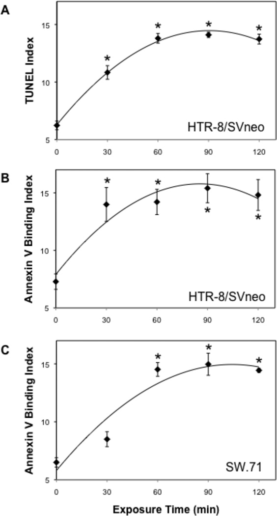
Background: Apoptosis is induced by ethanol (EtOH) in human placental trophoblast cells, possibly disrupting placentation and contributing to intrauterine growth restriction in fetal alcohol spectrum disorder (FASD). EtOH induces programmed cell death in several embryonic tissues by raising intracellular Ca(2+) . Therefore, the role of Ca(2+) signaling in EtOH-induced apoptosis was examined using human first trimester cytotrophoblast cell lines, examining the hypothesis that apoptosis is dependent on intracellular Ca(2+) signaling.
Methods: Using HTR-8/SVneo and SW.71 cytotrophoblast cell lines, real-time intracellular Ca(2+) concentration was monitored by fluo-4 epifluorescence microscopy and apoptosis was assessed by flow cytometry of cells fluorescently labeled for DNA fragmentation (TUNEL) and annexin V binding.
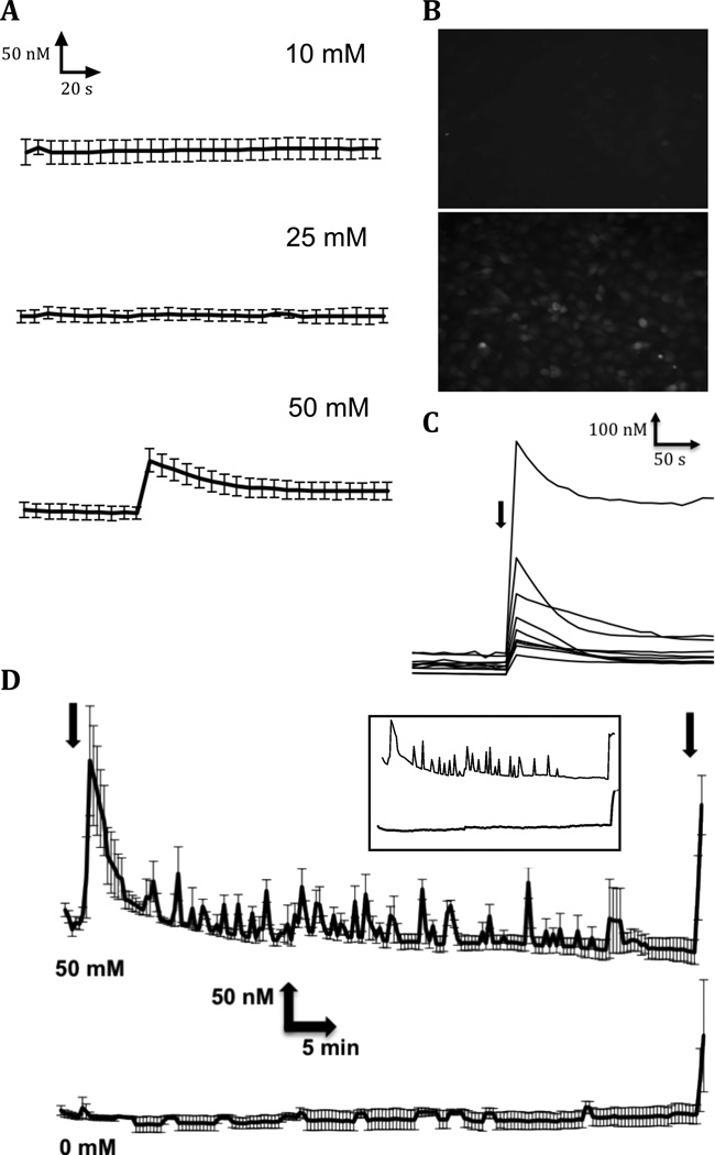
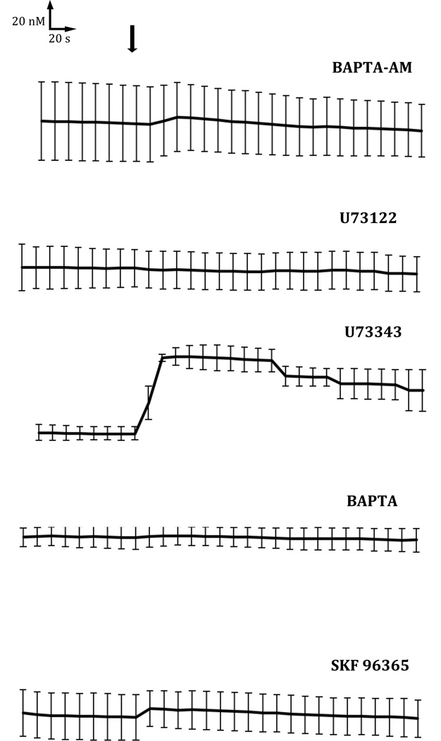
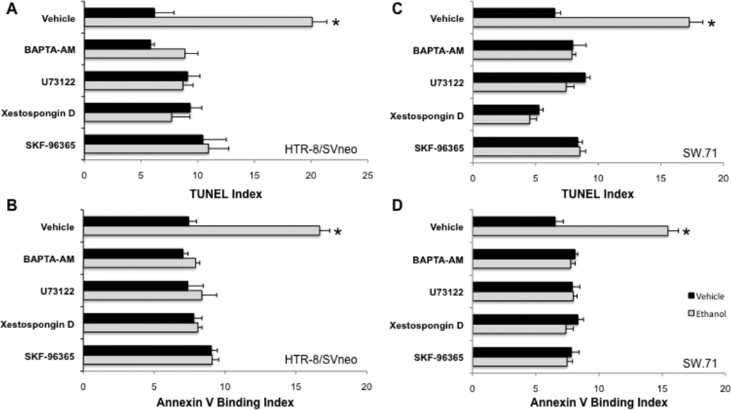
Results: Intracellular Ca(2+) concentrations increased synchronously in all cells within 10 seconds of exposure to 50 mM EtOH, but not at lower EtOH concentrations (10 to 25 mM) incapable of inducing apoptosis. Trophoblast cells treated with inhibitors of Ca(2+) signaling (BAPTA-AM, U73122, xestospongin D, BAPTA, SKF-96365) produced no intracellular Ca(2+) transients after exposure to 50 mM EtOH and were protected from cell death induced by EtOH.
Conclusions: EtOH-induced apoptosis in human cytotrophoblast cells, identified by DNA fragmentation and externalized phosphatidylserine, was dependent upon Ca(2+) signaling. Both intracellular Ca(2+) mobilization and extracellular Ca(2+) influx were required, as well as phosphatidylinositol signaling. Inhibition by SKF-96365 suggests that the capacitative Ca(2+) entry mechanism that utilizes TRPC channels was activated by EtOH. Apoptosis occurs downstream of Ca(2+) signaling in trophoblasts and may contribute to placental insufficiency and poor fetal growth associated with FASD.
Keywords: Alcohol; Apoptosis; Calcium; Fetal Alcohol Spectrum Disorder; Intracellular Signaling; Intrauterine Growth Restriction; Trophoblast.
Copyright © 2014 by the Research Society on Alcoholism.
Figures
Similar articles
-
Nifedipine Prevents Apoptosis of Alcohol-Exposed First-Trimester Trophoblast Cells.Alcohol Clin Exp Res. 2018 Jan;42(1):53-60. doi: 10.1111/acer.13534. Epub 2017 Nov 22. Alcohol Clin Exp Res. 2018. PMID: 29048755 Free PMC article.
-
Enhancement of trophoblast differentiation and survival by low molecular weight heparin requires heparin-binding EGF-like growth factor.Hum Reprod. 2017 Jun 1;32(6):1218-1229. doi: 10.1093/humrep/dex069. Hum Reprod. 2017. PMID: 28402449 Free PMC article.
-
Epidermal growth factor-like growth factors prevent apoptosis of alcohol-exposed human placental cytotrophoblast cells.Biol Reprod. 2007 Jul;77(1):53-60. doi: 10.1095/biolreprod.106.057984. Epub 2007 Mar 28. Biol Reprod. 2007. PMID: 17392498 Free PMC article.
-
Involvement of mTOR, JNK and PI3K in the negative effect of ethanol and metformin on the human first-trimester extravillous trophoblast HTR-8/SVneo cell line.Eur J Pharmacol. 2018 Aug 15;833:16-24. doi: 10.1016/j.ejphar.2018.05.038. Epub 2018 May 25. Eur J Pharmacol. 2018. PMID: 29807029
-
Detrimental effects of ethanol and its metabolite acetaldehyde, on first trimester human placental cell turnover and function.PLoS One. 2014 Feb 4;9(2):e87328. doi: 10.1371/journal.pone.0087328. eCollection 2014. PLoS One. 2014. PMID: 24503565 Free PMC article.
Cited by
-
Chronic ethanol exposure induces SK-N-SH cell apoptosis by increasing N-methyl-D-aspartic acid receptor expression and intracellular calcium.Exp Ther Med. 2018 Apr;15(4):3791-3800. doi: 10.3892/etm.2018.5902. Epub 2018 Feb 28. Exp Ther Med. 2018. PMID: 29581737 Free PMC article.
-
Nifedipine Prevents Apoptosis of Alcohol-Exposed First-Trimester Trophoblast Cells.Alcohol Clin Exp Res. 2018 Jan;42(1):53-60. doi: 10.1111/acer.13534. Epub 2017 Nov 22. Alcohol Clin Exp Res. 2018. PMID: 29048755 Free PMC article.
-
TRPV1 Contributes to the Neuroprotective Effect of Dexmedetomidine in Pilocarpine-Induced Status Epilepticus Juvenile Rats.Biomed Res Int. 2020 Apr 7;2020:7623635. doi: 10.1155/2020/7623635. eCollection 2020. Biomed Res Int. 2020. PMID: 32337274 Free PMC article.
-
The Placenta as a Target for Alcohol During Pregnancy: The Close Relation with IGFs Signaling Pathway.Rev Physiol Biochem Pharmacol. 2021;180:119-153. doi: 10.1007/112_2021_58. Rev Physiol Biochem Pharmacol. 2021. PMID: 34159446 Review.
-
Bias and misleading concepts in an Arnica research study. Comments to improve experimental Homeopathy.J Ayurveda Integr Med. 2018 Jan-Mar;9(1):75-80. doi: 10.1016/j.jaim.2017.01.014. Epub 2018 Feb 26. J Ayurveda Integr Med. 2018. PMID: 29496319 Free PMC article. Review.
References
-
- Axt R, Kordina AC, Meyberg R, Reitnauer K, Mink D, Schmidt W. Immunohistochemical evaluation of apoptosis in placentae from normal and intrauterine growth-restricted pregnancies. Clin.Exp.Obstet.Gynecol. 1999;26:195–198. - PubMed
-
- Barrio E, Calvo MT, Romo A, Alvarez R, Gutierrez JI, Naval J, Ferrandez LA. Intrauterine growth retardation: study of placental apoptosis. J.Pediatr.Endocrinol.Metab. 2004;17(Suppl 3):451–456. - PubMed
-
- Carvou N, Norden AG, Unwin RJ, Cockcroft S. Signalling through phospholipase C interferes with clathrin-mediated endocytosis. Cell Signal. 2007;19:42–51. - PubMed
-
- De A, Boyadjieva NI, Sarkar DK. Effect of voltage-dependent calcium channel blockers on ethanol-induced beta-endorphin release from hypothalamic neurons in primary cultures. Alcohol Clin Exp Res. 1999;23:850–855. - PubMed
-
- Debelak-Kragtorp KA, Armant DR, Smith SM. Ethanol-induced cephalic apoptosis requires phospholipase C-dependent intracellular calcium signaling. Alcohol Clin Exp Res. 2003;27:515–523. - PubMed
Publication types
MeSH terms
Substances
Grants and funding
LinkOut - more resources
Full Text Sources
Other Literature Sources
Miscellaneous
