Review
. 2014 Jul;37(7):370-80.
doi: 10.1016/j.tins.2014.05.001. Epub 2014 Jun 2.

How the brainstem controls orofacial behaviors comprised of rhythmic actions

Affiliations
Review

How the brainstem controls orofacial behaviors comprised of rhythmic actions

Jeffrey D Moore et al. Trends Neurosci. 2014 Jul.

Abstract

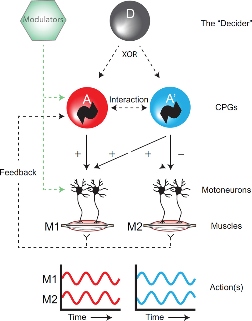
Mammals perform a multitude of well-coordinated orofacial behaviors such as breathing, sniffing, chewing, licking, swallowing, vocalizing, and in rodents, whisking. The coordination of these actions must occur without fault to prevent fatal blockages of the airway. Deciphering the neuronal circuitry that controls even a single action requires understanding the integration of sensory feedback and executive commands. A far greater challenge is to understand the coordination of multiple actions. Here, we focus on brainstem circuits that drive rhythmic orofacial actions. We discuss three neural computational mechanisms that may enable circuits for different actions to operate without interfering with each other. We conclude with proposed experimental programs for delineating the neural control principles that have evolved to coordinate orofacial behaviors.

Keywords: brainstem; central pattern generator; orofacial movements; pre-Bötzinger complex; vibrissa.


Figures

Figure 1
Figure 1. Schematic of the possible circuit arrangements for execution of different actions using a shared motor plant
Muscles M1 and M2 can both be used in different temporal patterns in two different actions, A and A’. Possible circuit interactions include: (1) CPGs interact and coordinate each other, (2) higher order centers (D) gate, or “select” separate CPGs, and (3) peripheral feedback into a CPG alters the phase relationship between the muscles. Additionally, various neuromodulators may act on either the CPGs themselves or their outputs to affect their frequency or amplitude.
Figure 2
Figure 2. Coordination between breathing and other rhythmic orofacial actions
(a) An isolated brainstem preparation in which rhythmic bursts of fictive motor activity were induced via bath application of NMDA (left). Hypoglossal and phrenic motor outputs were monitored electrophysiologically via the XIIth cranial rootlet and the 5th cervical rootlet, respectively (black traces, right). The integrated activity of the XIIth rootlet is shown in green. Phrenic bursts are reported to reset the phase of the faster hypoglossal rhythm. Adapted from, . (b) Simultaneous monitoring of licking (green) and breathing (black) in an alert rat (left and middle) show that the actions are coordinated (right). The occurrence of a lick is dependent on the phase of the respiratory cycle. Adapted from. (c) Simultaneous monitoring of whisking (red) and breathing (black) in an alert rat (left and middle) show that the actions are coordinated (right). Protraction and inspiration are upward. Inspiration is synchronous with protraction on each cycle (top middle) during sniffing but only with a fraction of the cycles during basal respiration (bottom middle), as intervening whisks occur. Rasters of inspiration onset (black) and protraction onset (red) times relative to the onset of inspiration for individual breath are ordered by the duration of the breath (right). At high respiratory rates, whisking and breathing show a 1:1 temporal relationship, while at lower breathing rates there are additional, intervening whisks between each breath. Adapted from. (d) Simultaneous monitoring of chewing (orange), swallowing (purple), and breathing (black) in an alert rabbit (left and middle) reveal the nature of their coordination. While breathing and chewing appear to be asynchronous, swallowing affects both rhythms. The occurrence of a swallowing movement delays subsequent breathing and chewing cycles. Adapted from.
Figure 3
Figure 3. Anatomy of neural circuits involved in generating and coordinating orofacial actions
(a) Three-dimensional reconstruction of the pons and medulla, which contain regions that receive primary somatosensory inputs. Cutaneous inputs from the face innervate the trigeminal sensory nuclei (blue). Proprioceptive innervation of the jaw muscles arises from cells in the trigeminal mesencephalic nucleus (pink). Gustatory inputs from the tongue innervate the solitary nucleus (NTS). The structure is shown in the sagittal (left), horizontal (middle) and frontal (right planes). Light transparent structures correspond to the motor nuclei in panel b. (b) The same reconstruction as in panel a, showing the pools of cranial motoneurons that control the jaws (orange), face (red), airway (yellow), and tongue (green). Conventions are as in panel a. Light transparent structures correspond to the sensory nuclei in panel a. (c) The same reconstruction as in panels a and b, showing the approximate locations of known premotor nuclei to each of the motoneuron pools in panel a. Premotor nuclei are color coded according to the primary motor nucleus that they innervate. The brainstem is shown in the sagittal (left) and frontal (right) planes. Breathing-related regions are shown in black. Abbreviations are as follows: parvocellular reticular formation (PCRt), caudal/rostral ventral respiratory groups (cVRG and rVRG, respectively), hypoglossal/vibrissa/trigeminal intermediate reticular formation (hIRt, vIRt, tIRt, respectively), preBotzinger complex (Pre-BotC), parafacial respiratory group (pFRG), gigantocellular reticular formation (Gi), lateral paragigantocellular reticular formation (LPGi), and dorsal principal trigeminal nucleus (dPrV). (d) The same reconstruction as in panels a–c, highlighting the locations of the putative neuronal oscillators (marked as “~”) that generate breathing (black), whisking (red), licking (green) and chewing (orange). Conventions are as in panels a–c. The location of the chewing oscillator remains unresolved.

References

    1. Weiss P. Self-differentiation of the basic patterns of coordination. Williams & Wilkins; 1941.
    1. Tinbergen N. The Study of Instinct. 1951
    1. Mussa–Ivaldi FA, Bizzi E. Motor learning through the combination of primitives. Philosophical Transactions of the Royal Society of London. Series B: Biological Sciences. 2000;355:1755–1769. - PMC - PubMed
    1. Sherrey JH, Megirian D. State dependence of upper airway respiratory motoneurons: Functions of the cricothyroid and nasolabial muscles of the unanesthetized rat. Electroencephalography and Clinical Neurophysiology. 1977;43:218–228. - PubMed
    1. Welker WI. Analysis of sniffing of the albino rat. Behaviour. 1964;12:223–244. (1964)

Publication types