. 2014 Mar-Apr;4(2):82-89.
doi: 10.1016/j.prro.2013.06.004. Epub 2013 Aug 7.

Upper abdominal normal organ contouring guidelines and atlas: a Radiation Therapy Oncology Group consensus

Affiliations

Upper abdominal normal organ contouring guidelines and atlas: a Radiation Therapy Oncology Group consensus

Salma K Jabbour et al. Pract Radiat Oncol. 2014 Mar-Apr.

Abstract

Purpose: To standardize upper abdominal normal organ contouring guidelines for Radiation Therapy Oncology Group (RTOG) trials.

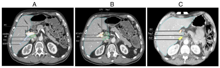
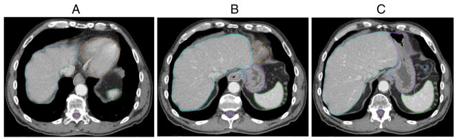
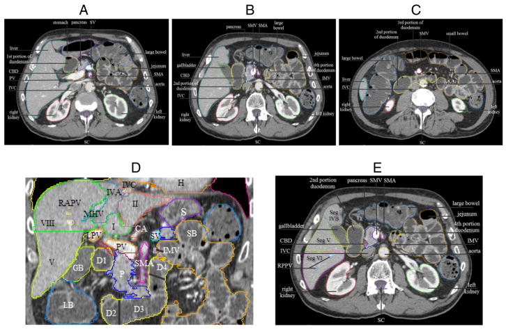
Methods and materials: Twelve expert radiation oncologists contoured the liver, esophagus, gastroesophageal junction (GEJ), stomach, duodenum, and common bile duct (CBD), and reviewed and edited 33 additional normal organ and blood vessel contours on an anonymized patient computed tomography (CT) dataset. Contours were overlaid and compared for agreement using MATLAB (MathWorks, Natick, MA). S95 contours, defined as the binomial distribution to generate 95% group consensus contours, and normal organ contouring definitions were generated and reviewed by the panel.

Results: There was excellent consistency and agreement of the liver, duodenal, and stomach contours, with substantial consistency for the esophagus contour, and moderate consistency for the GEJ and CBD contours using a Kappa statistic. Consensus definitions, detailed normal organ contouring recommendations and high-resolution images were developed.

Conclusions: Consensus contouring guidelines and a CT image atlas should improve contouring uniformity in radiation oncology clinical planning and RTOG trials.


Conflict of interest statement

Conflicts of interest: None.

Figures

Figure 1
Figure 1
For purposes of contouring the liver, the portal vein should be included in the liver (light blue) contour when segment (Seg) I (caudate lobe) is left of the portal vein (PV). (A) Demonstrates segment I, “caudate tail,” posterior to the PV, so the liver contour should exclude the PV. (B) Demonstrates segment I to the left of the PV, so the liver contour should include segment I and the PV. Left portal vein (LPV), right anterior portal vein (RTAPV), inferior vena cava (IVC), Right posterior portal vein (RPPV) in (A). (C) Demonstrates a patient example in which segment I does not fully extend to the left of the PV, and the PV is excluded from the liver contour.
Figure 2
Figure 2
(A) Consensus contours of liver (light blue), inferior vena cava (IVC) (yellow), esophagus (white), heart (orange), spleen (green), stomach (purple in 2B and 2C), spinal canal (purple), top of gastroesophageal junction (GEJ) (light blue in 2B), and bottom of GEJ (light blue in 2C), large bowel (2C).
Figure 3
Figure 3
(A), (B), and (C) Axial images demonstrating 4 portions of the duodenum (shown in yellow). Splenic vein (SV), portal vein (PV), common bile duct (CBD), inferior vena cava (IVC), superior mesenteric artery (SMA), SMV, spinal canal (SC). (D) Coronal images illustrating the 4 portions of duodenum and liver segments. Liver segments I, II, IVA, V, VIII, and D1–D4: first to fourth portions of duodenum. IMV, inferior mesenteric vein, demarcates the duodenojejunal junction; RAPV, right anterior portal vein; MHV, middle hepatic vein; LPV, left portal vein; IVC (yellow) with adjacent left hepatic vein (blue, not labeled); H, heart; S, stomach; LB, large bowel; SB, small bowel; GB, gallbladder; P, pancreas; PV, portal vein, to the right of the PV is CBD (red), to the right and inferior of the PV is superior mesenteric vein (SMV, burgundy) (ie, SMV-PV confluence); CA, celiac artery, with splenic artery (orange) to the right and inferior of CA. (E) Axial image showing duodenojejunal junction demarcated by IMV.
Figure 4
Figure 4
Liver segments (Seg), demonstrated on axial images, descending from superior to inferior slices (A)–(E). CA, celiac axis; LVH, left hepatic vein; MHV, middle hepatic vein; RHV, right hepatic vein; RPPV, right posterior portal vein; RTAPV, right anterior portal vein; LPV, left portal vein; SA, splenic artery; IVC, inferior vena cava; CHA, common hepatic artery; SV, splenic vein; SC, spinal canal.

References

    1. Pan H, Simpson DR, Mell LK, Mundt AJ, Lawson JD. A survey of stereotactic body radiotherapy use in the United States. Cancer. 2011;117:4566–4572. - PMC - PubMed
    1. Gay HA, Barthold HJ, O’Meara E, et al. Pelvic normal tissue contouring guidelines for radiation therapy: a Radiation Therapy Oncology Group consensus panel atlas. Int J Radiat Oncol Biol Phys. 2012;83:e353–e362. - PMC - PubMed
    1. Kong FM, Ritter T, Quint DJ, et al. Consideration of dose limits for organs at risk of thoracic radiotherapy: atlas for lung, proximal bronchial tree, esophagus, spinal cord, ribs, and brachial plexus. Int J Radiat Oncol Biol Phys. 2011;81:1442–1457. - PMC - PubMed
    1. Goodman KA, Regine WF, Dawson LA, et al. Radiation Therapy Oncology Group consensus panel guidelines for the delineation of the clinical target volume in the postoperative treatment of pancreatic head cancer. Int J Radiat Oncol Biol Phys. 2012;83:901–908. - PMC - PubMed
    1. Deasy JO, Blanco AI, Clark VH. CERR: a computational environment for radiotherapy research. Med Phys. 2003;30:979–985. - PubMed

Publication types

MeSH terms