Review
. 2014 Jun 2;6(6):a021857.
doi: 10.1101/cshperspect.a021857.

CSF-1 receptor signaling in myeloid cells

Affiliations
Review

CSF-1 receptor signaling in myeloid cells

E Richard Stanley et al. Cold Spring Harb Perspect Biol. .

Abstract

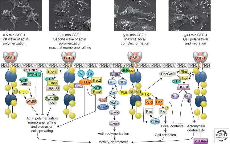
The CSF-1 receptor (CSF-1R) is activated by the homodimeric growth factors colony-stimulating factor-1 (CSF-1) and interleukin-34 (IL-34). It plays important roles in development and in innate immunity by regulating the development of most tissue macrophages and osteoclasts, of Langerhans cells of the skin, of Paneth cells of the small intestine, and of brain microglia. It also regulates the differentiation of neural progenitor cells and controls functions of oocytes and trophoblastic cells in the female reproductive tract. Owing to this broad tissue expression pattern, it plays a central role in neoplastic, inflammatory, and neurological diseases. In this review we summarize the evolution, structure, and regulation of expression of the CSF-1R gene. We discuss the structures of CSF-1, IL-34, and the CSF-1R and the mechanism of ligand binding to and activation of the receptor. We further describe the pathways regulating macrophage survival, proliferation, differentiation, and chemotaxis downstream from the CSF-1R.


Figures

Figure 1.
Figure 1.
Structure of the CSF-1R and regulation of Csf1r gene expression. (A) Structures of CSF-1R and oncogenic derivatives. (Left) The c-fms proto-oncogene, (middle) the v-fms oncogene, encoded by the Susan McDonough strain of feline sarcoma virus (SM-FeSV), and (right) the CSF-1R–RBL6 oncogenic fusion protein. Ovals D1–D5 represent the five extracellular Ig-like domains of CSF-1R. The ligand-binding domains are gray. The blue dots in D4 represent the ionic pairs that have been implicated in receptor homotypic contacts. The intracellular domain is shown as the juxtamembrane domain (JMD, orange), kinase N lobe (ATP, dark blue), kinase insert (KI, green), kinase C lobe (Kin, light blue), activation loop (AL, purple), and carboxy-terminal tail (black). All amino acid substitutions in v-fms are shown as annotated red dots and the carboxy-terminal amino acid sequence that is unrelated to c-fms is red (Woolford et al. 1988). (B) Evolution of closely related type III RTK genes by gene and genome duplications (based on data from Braasch et al. 2006). (C) (Top) Exon-intron structure of mouse Csf1r gene and (bottom) expanded promoter structure and transcription factor (TF) binding sites (based on data from Bonifer and Hume 2008; Ovchinnikov et al. 2010). (D) Regulation of Csf1r expression in hematopoiesis (based on data from Bonifer and Hume 2008). The silenced state levels for each parameter were measured in T cells and fibroblasts that do not express the CSF-1R. Mo, monocyte; Mϕ, macrophage.
Figure 2.
Figure 2.
Structure of CSF-1, IL-34, and CSF-1R ligand–receptor complexes. (A) Topological diagrams of monomeric (left) and ribbon representations of dimeric CSF-1 (top) and IL-34 (bottom) (based on data from Pandit et al. 1992; Liu et al. 2012; Ma et al. 2012). Gray lines in topological diagrams represent intramolecular disulfide bridges; dotted gray line in CSF-1 indicates position of the intermolecular disulfide bond. (B) Structure of CSF-1R ectodomain complex with IL-34 and CSF-1 (based on data from Felix et al. 2013). (C) Model of early events in CSF-1R activation (based on data from Verstraete and Savvides 2012). Gold spheres, phosphotyrosines (P), gray spheres, ubiquitination (Ub). Note that phosphorylation of CSF-1R tyrosine residue 559 and intracellular domain ubiquitination are important for full receptor activation and tyrosine phosphorylation.
Figure 3.
Figure 3.
CSF-1R signaling in macrophage survival and proliferation. (A) Biological functions regulated by individual CSF-1R tyrosine residues. (B) Protein interactions and signaling events triggered by individual CSF-1R phosphotyrosine residues (based on data from Pixley and Stanley 2004). (C) CSF-1R signaling for macrophage survival. (D) Pathways mediating CSF-1R proliferative responses in macrophages. Arrows indicate activation; black line-capped arrows, inhibition; gray line-capped arrows, late-phase inhibition; round-capped arrows, increased expression or concentration; diamond-capped arrows, dissociation; dotted arrows, partial contribution; gold spheres, phosphotyrosines; and silver spheres, serine/threonine phosphorylation. Numbered gold spheres indicate the mouse CSF-1R tyrosine residues required for activation of specific pathways. Those without numbers indicate that the CSF-1R phosphotyrosyl residue triggering the response is not known.
Figure 4.
Figure 4.
CSF-1R signaling in macrophage differentiation. (Top) Morphology of May-Grünwald-Giemsa-stained macrophage precursors. Gray arrows (middle panel) indicate that Gab2 significantly contributes to these pathways in monoblasts, but not in pro-monocytes. Symbols are as described in the legend for Figure 3.
Figure 5.
Figure 5.
CSF-1R signaling in macrophage migration and chemotaxis. (Top panels) Scanning electron microscopic images of macrophages stimulated with CSF-1 for the indicated times. The dotted gray arrow indicates a hypothetical mechanism. Symbols are as described in the legend for Figure 3.

References

    1. Abou-Kheir W, Isaac B, Yamaguchi H, Cox D 2008. Membrane targeting of WAVE2 is not sufficient for WAVE2-dependent actin polymerization: A role for IRSp53 in mediating the interaction between Rac and WAVE2. J Cell Sci 121: 379–390 - PMC - PubMed
    1. Allen WE, Zicha D, Ridley AJ, Jones GE 1998. A role for Cdc42 in macrophage chemotaxis. J Cell Biol 141: 1147–1157 - PMC - PubMed
    1. Baccarini M, Li W, Dello Sbarba P, Stanley ER 1991. Increased phosphorylation of the colony stimulating factor-1 receptor following transmembrane signaling. Receptor 1: 243–259 - PubMed
    1. Baran CP, Tridandapani S, Helgason CD, Humphries RK, Krystal G, Marsh CB 2003. The inositol 5′-phosphatase SHIP-1 and the Src kinase Lyn negatively regulate macrophage colony-stimulating factor-induced Akt activity. J Biol Chem 278: 38628–38636 - PubMed
    1. Bartelmez SH, Stanley ER 1985. Synergism between hemopoietic growth factors (HGFs) detected by their effects on cells bearing receptors for a lineage specific HGF: Assay of hemopoietin-1. J Cell Physiol 122: 370–378 - PubMed

Publication types

MeSH terms

Substances