. 2014 Aug;105(8):933-42.
doi: 10.1111/cas.12459. Epub 2014 Aug 11.

Th17 cells and interleukin-17 increase with poor prognosis in patients with acute myeloid leukemia

Affiliations

Th17 cells and interleukin-17 increase with poor prognosis in patients with acute myeloid leukemia

Yixiang Han et al. Cancer Sci. 2014 Aug.

Abstract

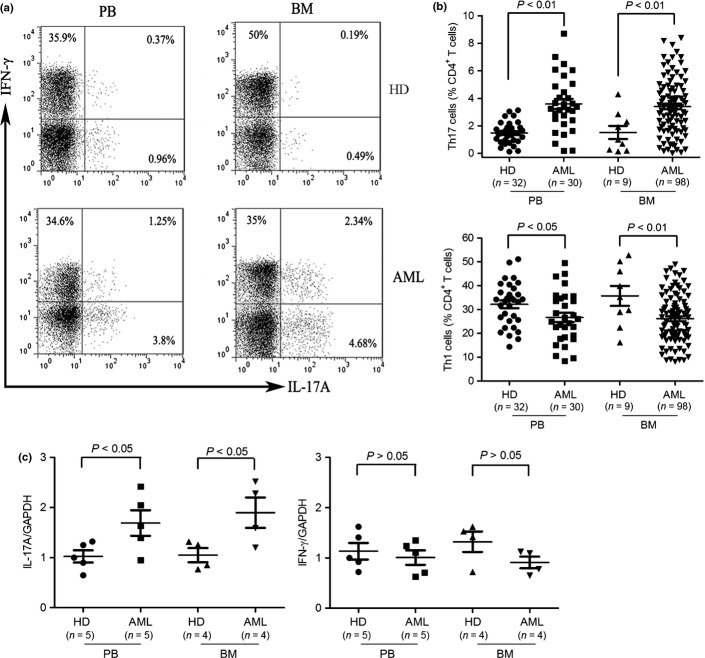
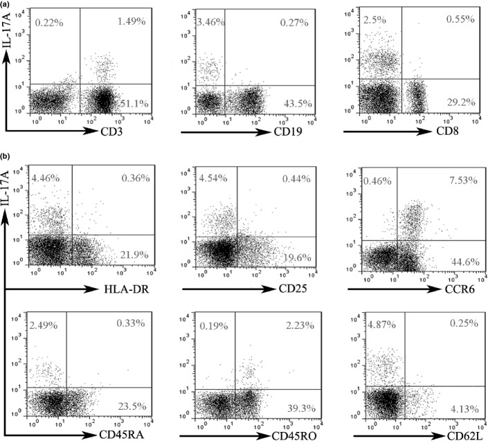
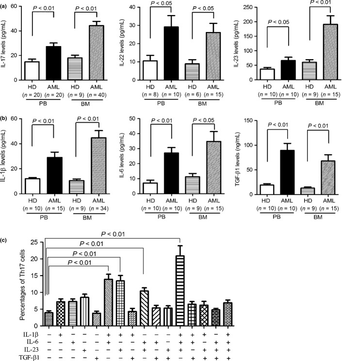
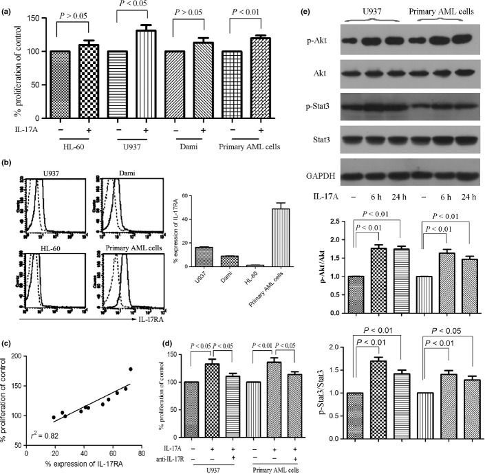
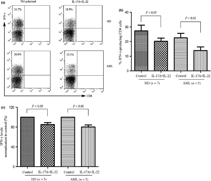
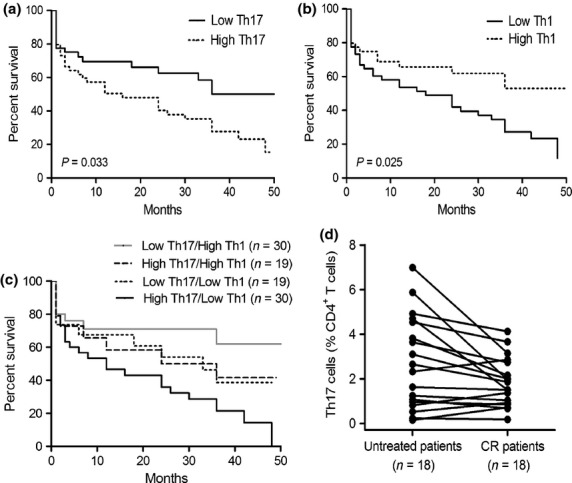
Although Th17 cells play crucial roles in the pathogenesis of many autoimmune and inflammatory disorders, their roles in malignancies are currently under debate. The role and mechanism of Th17 cells in patients with acute myeloid leukemia (AML) remain poorly understood. Here we demonstrated that the frequency of Th17 cells was significantly increased in peripheral blood mononuclear cells (PBMCs) and bone marrow mononuclear cells from AML patients compared with healthy donors. Plasma levels of interleukin (IL)-17, IL-22, IL-23, IL-1β, IL-6, and transforming growth factor (TGF)-β1 were significantly increased in blood and bone marrow in AML patients compared with healthy donors. The in vitro experiments demonstrated that IL-1β, IL-6, IL-23, but not TGF-β1 promoted the generation and differentiation of Th17 cells from naive CD4(+) T cells in humans. IL-17A, a signature cytokine secreted by Th17 cells, induced the proliferation of IL-17 receptor (IL-17R)-positive AML cells via IL-17R, in which activation of PI3K/Akt and Jak/Stat3 signaling pathway may play important roles. In addition, combination of IL-17A and IL-22 significantly reduced the generation of Th1 cells and the production of interferon (IFN)-γ from healthy donor or AML patient peripheral blood mononuclear cells. Patients with high Th17 cell frequency had poor prognosis, whereas patients with high Th1 cell frequency had prolonged survival. Combined analysis of Th1 and Th17 cell frequencies improved the ability to predict patient outcomes. In conclusion, Th17 cells play a crucial role in the pathogenesis of AML and may be an important therapeutic target and prognostic predictor.

Keywords: Acute myeloid leukemia; Th1 cells; Th17 cells; interleukin-17; prognostic factors.


Figures

Fig. 1
Fig. 1
Elevated frequencies of Th17 cells and reduced frequencies of Th1 cells in freshly isolated peripheral blood mononuclear cells (PBMCs) and bone marrow mononuclear cells (BMMCs) from acute myeloid leukemia (AML) patients. (a) PBMCs and BMMCs were isolated from AML patients and healthy donors (HDs) and stimulated for 5 h with phorbol 12-myristate13-acetate (PMA) and ionomycin in the presence of brefeldin A and then stained for CD3, CD8, intracellular interleukin (IL)-17A and interferon (IFN)-γ. Frequencies of Th17 cells and Th1 cells were determined by flow cytometry. Representative dot plots using matching peripheral blood (PB) and bone marrow (BM) samples from AML patients and HDs were shown. (b) Collective results presented for Th17 and Th1 cells within CD4+ T population. (c) Total RNA was isolated from CD4+ T cells obtained from AML patients and HDs and reverse transcribed into cDNA and subsequently real time polymerase chain reaction (PCR) for IL-17A and IFN-γ. Results were expressed as mean ± SEM.
Fig. 2
Fig. 2
Phenotype of tumor-infiltrating Th17 cells. After stimulation with phorbol 12-myristate13-acetate (PMA) and ionomycin for 5 h, freshly isolated bone marrow mononuclear cells (BMMCs) were subjected to membrane and intracellular staining and analyzed by flow cytometry. Representative data were shown from 21 untreated AML patients. (a) Interleukin (IL)-17A expression in T and B cells. IL-17A expression was analyzed in BMMCs. (b) The expression of HLA-DR, CD25, CCR6, CD45RA, CD45RO, and CD62L in tumor-infiltrating Th17 cells.
Fig. 3
Fig. 3
Generation and differentiation of Th17 cells from peripheral blood (PB) and bone marrow (BM) regulated by interleukin (IL)-1β, IL-6, and IL-23. Elevated levels of Th17-producing cytokines (a) and Th17-associated proinflammatory cytokines (b) in PB and BM samples from acute myeloid leukemia (AML) patients were determined by ELISA. (c) Th17 cells detected in naive CD4+ T cells purified from AML patients after stimulation with indicated cytokines. The naive CD4+ T cells were isolated from PB of AML patients and subsequently stimulated by plate-bound anti-CD3 and soluble anti-CD28 mAbs with or without the indicated cytokines. After 7 days, the cells were restimulated with phorbol 12-myristate13-acetate (PMA) and ionomycin in the presence of brefeldin A and analyzed for intercellular IL-17A expression. The data were expressed as mean ± SEM representing four independent experiments from different AML patients.
Fig. 4
Fig. 4
Interleukin (IL)-17A promotes the proliferation of AML cells via IL-17 receptor (IL-17R). (a) Acute myeloid leukemia (AML) cell lines HL-60, U937, Dami, and primary AML cells isolated from AML patients (n = 23) were incubated with or without IL-17A (50 ng/mL) for 7 days and proliferation was assayed by MTT. Data are showed in proliferation in the presence of IL-17A compared with control and expressed as mean ± SEM representing at least three independent experiments. (b) The IL-17RA expression of AML cells was measured using anti-CD217 PE antibody (solid line) or mouse IgG1 PE antibody (dotted line) by flow cytometry. Representative histograms (left panel) and statistical data (right panel) were shown. (c) A correlation was observed between the IL-17RA expression and the IL-17A-inducing proliferation. (d) The effects of IL-17R on U937 and primary AML cell proliferation were determined by incubating the cells with IL-17A (50 ng/mL) in the presence or absence of anti-IL-17R antibody (3 μg/mL) for 7 days. (e) Western blotting showed the phosphorylation of Akt and Stat3 were significantly increased after IL-17A stimulation for 6 h and lasted for 24 h in IL-17R+ AML cells. Representatives and statistical data were shown for four independent experiments.
Fig. 5
Fig. 5
Interleukin (IL)-17A and IL-22 inhibited IL-12-inducing interferon (IFN)-γ-producing cells in peripheral blood mononuclear cells (PBMCs). (a) & (b) PBMCs from healthy donors or AML patients were activated with Th1 polarizing cytokines (IL-12, 10 ng/mL; anti-CD3 antibody, 1.5 μg/mL) with or without IL-17A (10 ng/mL) and IL-22 (10 ng/mL) for 12 days. Cells were subsequently stimulated with phorbol 12-myristate13-acetate (PMA) and ionomycin in the presence of brefeldin A, stained for intercellular IFN-γ, and analyzed by flow cytometry. A representative dot plot analysis showing percentage of IFN-γ-positive cells within gated CD3+CD4+ (CD3+CD8-) population. Results are expressed as mean ± SEM representing five independent experiments from different healthy donors or AML patients. (c) PBMCs from healthy donors or AML patients were stimulated with Th1 polarizing cytokines with or without IL-17A and IL-22 for 6 days, and supernatants were determined for IFN-γ by ELISA.
Fig. 6
Fig. 6
Increased frequencies of Th17 cells and reduced frequencies of Th1 cells predict poor survival in acute myeloid leukemia (AML) patients. Kaplan–Meier curves for overall survival were assessed in combination with frequencies of Th17 cells (a), frequencies of Th1 cells (b), or frequencies of Th1 together with Th17 cells (c), in bone marrow (BM) microenvironment of 98 AML patients. (d) The frequencies of Th17 cells decreased in BM when patients achieved complete remission (CR).

References

    1. Le Dieu R, Taussig DC, Ramsay AG, et al. Peripheral blood T cells in acute myeloid leukemia (AML) patients at diagnosis have abnormal phenotype and genotype and form defective immune synapses with AML blasts. Blood. 2009;114:3909–16. - PMC - PubMed
    1. Ustun C, Miller JS, Munn DH, Weisdorf DJ, Blazar BR. Regulatory T cells in acute myelogenous leukemia: is it time for immunomodulation? Blood. 2011;118:5084–95. - PMC - PubMed
    1. Korn T, Bettelli E, Oukka M, Kuchroo VK. IL-17 and Th17 Cells. Annu Rev Immunol. 2009;27:485–517. - PubMed
    1. Ouyang W, Kolls JK, Zheng Y. The biological functions of T helper 17 cell effector cytokines in inflammation. Immunity. 2008;28:454–67. - PMC - PubMed
    1. Huber M, Heink S, Pagenstecher A, et al. IL-17A secretion by CD8+ T cells supports Th17-mediated autoimmune encephalomyelitis. J Clin Invest. 2013;123:247–60. - PMC - PubMed

Publication types

MeSH terms