Clinical Trial
. 2014 Jul 1;193(1):41-7.
doi: 10.4049/jimmunol.1301753. Epub 2014 Jun 2.

Aspirin activation of eosinophils and mast cells: implications in the pathogenesis of aspirin-exacerbated respiratory disease

Affiliations
Clinical Trial

Aspirin activation of eosinophils and mast cells: implications in the pathogenesis of aspirin-exacerbated respiratory disease

John W Steinke et al. J Immunol. .

Abstract

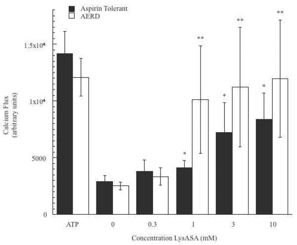
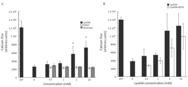
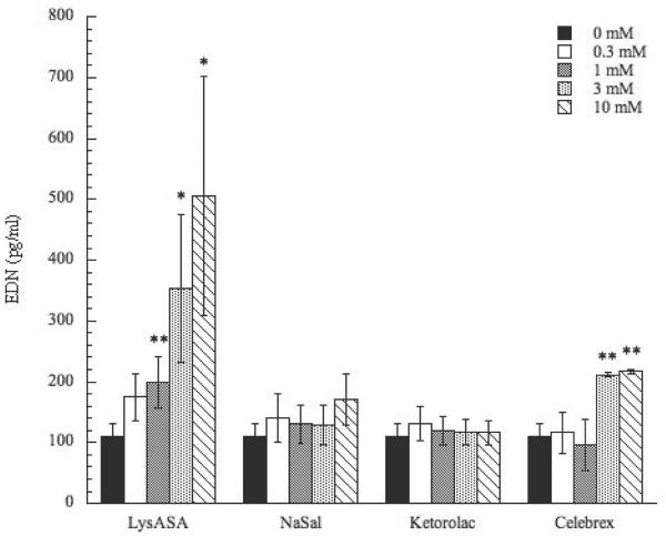
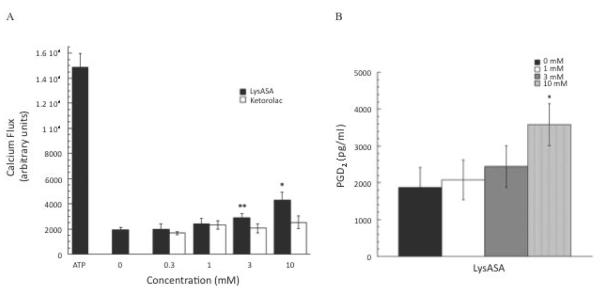
Reactions to aspirin and nonsteroidal anti-inflammatory drugs in patients with aspirin-exacerbated respiratory disease (AERD) are triggered when constraints upon activated eosinophils, normally supplied by PGE2, are removed secondary to cyclooxygenase-1 inhibition. However, the mechanism driving the concomitant cellular activation is unknown. We investigated the capacity of aspirin itself to provide this activation signal. Eosinophils were enriched from peripheral blood samples and activated with lysine ASA (LysASA). Parallel samples were stimulated with related nonsteroidal anti-inflammatory drugs. Activation was evaluated as Ca2+ flux, secretion of cysteinyl leukotrienes (CysLT), and eosinophil-derived neurotoxin (EDN) release. CD34+ progenitor-derived mast cells were also used to test the influence of aspirin on human mast cells with measurements of Ca2+ flux and PGD2 release. LysASA induced Ca2+ fluxes and EDN release, but not CysLT secretion from circulating eosinophils. There was no difference in the sensitivity or extent of activation between AERD and control subjects, and sodium salicylate was without effect. Like eosinophils, aspirin was able to activate human mast cells directly through Ca2+ flux and PGD2 release. AERD is associated with eosinophils maturing locally in a high IFN-γ milieu. As such, in additional studies, eosinophil progenitors were differentiated in the presence of IFN-γ prior to activation with aspirin. Eosinophils matured in the presence of IFN-γ displayed robust secretion of both EDN and CysLTs. These studies identify aspirin as the trigger of eosinophil and mast cell activation in AERD, acting in synergy with its ability to release cells from the anti-inflammatory constraints of PGE2.


Figures

Figure 1
Figure 1. LyASA stimulation of eosinophils from AERD and non-AERD subjects
Calcium mobilization assays were performed on eosinophils by plating 125,000 cells/well and incubating the cells with Fluo-4 NW wash dye mix supplemented with 5mM probenecid for 1 hr. Dose-response curves were generated by incubation of the cells increasing concentrations of LysASA (0.3–10mM) and measuring cytoplasmic calcium flux. ATP was used as a positive control. Data are presented as the mean±SEM of separate studies performed in triplicate (6 aspirin tolerant, 6 AERD subjects: *p<0.05 and **p<0.05 compared to unstimulated healthy control and AERD control, respectively).
Figure 2
Figure 2. Ca+2 flux of NSAID stimulated eosinophils
Calcium mobilization assays were performed on eosinophils by plating 125,000 cells/well and incubating the cells with Fluo-4 NW wash dye mix supplemented with 5mM probenecid for 1 hr. A) Dose-response curves were generated by incubation of the cells increasing concentrations of LysASA, NaSal or ketorolac (0.3–10mM) and measuring cytoplasmic calcium flux. ATP was used as a positive control. Data are presented as the mean±SEM of separate studies performed in triplicate (n=9 LysASA and ketorolac, n=6 NaSal: *p<0.05 compared to unstimulated control). B) Dose-response curves were generated by incubation of the cells with increasing concentrations of LysASA (0.3–10mM) in the presence or absence 1mM EGTA and measuring cytoplasmic Ca+2 flux.
Figure 3
Figure 3. Activation of peripheral blood eosinophils by NSAIDs
Eosinophils were purified from peripheral blood and stimulated in a dose-dependent fashion with LysASA, NaSal, ketorolac, or celecoxib (0.3–10mM) for 30 min. EDN was measured in supernatants and measured in ng/ml. Data are presented as the mean±SEM of 4–13 separate studies. *p<0.05; **p<0.01 as compared to unstimulated cells.
Figure 4
Figure 4. Activation of eosinophils differentiated from hematopoietic precursors in the additional presence of IFN-γ
CD34+-enriched hematopoietic progenitor cells were cultured for 3 days with SCF, TPO, Flt3L, IL-3, and IL-5 after which they were cultured for 3 additional weeks with just IL-3 and IL-5 with or without the additional presence of IFN-γ. Eosinophils matured from CD34+ progenitors were activated with or without calcium ionophore/PMA, LysASA (1 and 10 mM) or ketorolac (1 and 10 mM) for 30 minutes. Supernatants were collected and A) EDN and B) CysLT levels quantified and measured in ng/ml or pg/ml, respectively. Data are presented as the mean±SEM of a minimum of 6 separate studies. ‡p=0.05, *p<0.0004, **p<0.03, ***p<0.002 as compared to unstimulated cells.
Figure 5
Figure 5. Activation of CD34-differentiated mast cells
A) Calcium mobilization assays were performed on CD34-derived mast cells as described. Dose-response curves were generated by incubation of the cells increasing concentrations of LysASA or ketorolac (0.3–10mM). ATP was used as a positive control. Data are presented as the mean±SEM of 3 separate studies performed in triplicate (*p<0.05, **p=0.08). B) CD34-derived mast cells were stimulated with LysASA (0.3–10mM) for 30 min. PGD2 release was measured in supernatants as pg/ml. Data are presented as the mean±SEM of 10 studies (p<0.03).

Similar articles

Cited by

References

    1. Steinke JW, Huyett P, Payne SC, Negri J, Borish L. Cytokines and lymphocytes in eosinophilic sinusitis: prominent role for interferon-γ. J Allergy Clin Immunol. 2011;132:856–865. - PMC - PubMed
    1. Mascia K, Borish L, Patrie J, Hunt J, Phillips CD, Steinke JW. Chronic hyperplastic eosiniphilic sinusistis as a predictor of aspirin-exacerbated respiratory disease. Ann Allergy Asthma Immunol. 2005;94:652–657. - PubMed
    1. Szczeklik A, Stevenson DD. Aspirin-induced asthma: Advances in pathogenesis and management. J Allergy Clin Immunol. 1999;104:5–13. - PubMed
    1. Szczeklik A, Nizankowska E. Clinical features and diagnosis of aspirin induced asthma. Thorax. 2000;55:S42–S44. - PMC - PubMed
    1. Berges-Gimeno MP, Simon RA, Stevenson DD. The natural history and clinical characteristics of aspirin-exacerbated respiratory disease. Ann Allergy Asthma Immunol. 2002;89:474–478. - PubMed

Publication types