. 2014 Jul 1;193(1):21-5.
doi: 10.4049/jimmunol.1400321. Epub 2014 Jun 2.

Cutting edge: Antigen-specific thymocyte feedback regulates homeostatic thymic conventional dendritic cell maturation

Affiliations

Cutting edge: Antigen-specific thymocyte feedback regulates homeostatic thymic conventional dendritic cell maturation

Nicholas A Spidale et al. J Immunol. .

Abstract

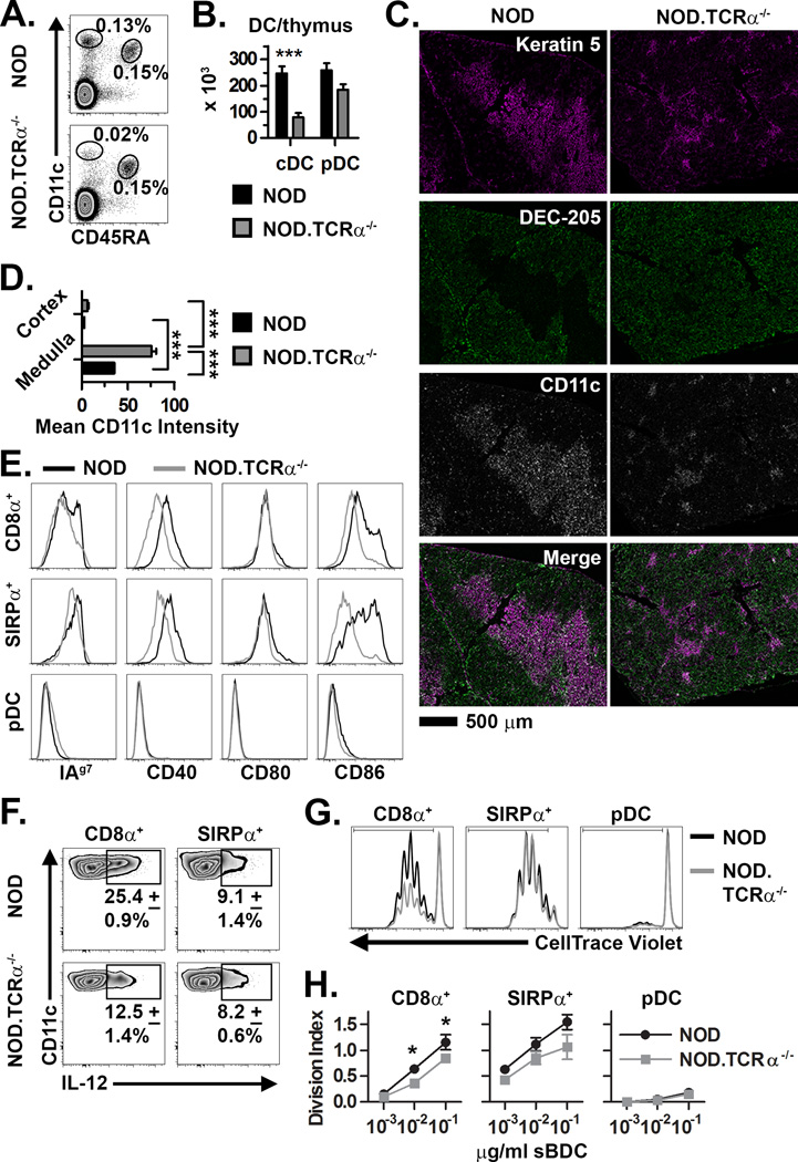
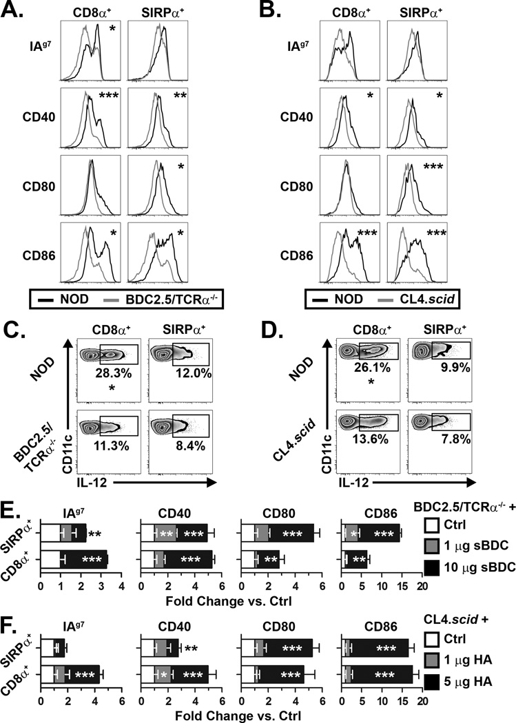
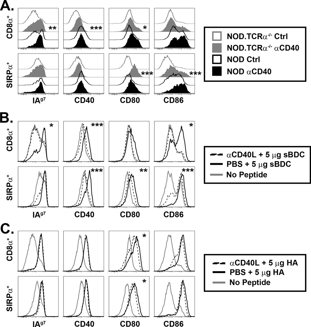
Thymic dendritic cells (DC) mediate self-tolerance by presenting self-peptides to and depleting autoreactive thymocytes. Despite a significant role in negative selection, the events regulating thymic DC maturation and function under steady-state conditions are poorly understood. We report that cross-talk with thymocytes regulates thymic conventional DC (cDC) numbers, phenotype, and function. In mice lacking TCR-expressing thymocytes, thymic cDC were reduced and exhibited a less mature phenotype. Furthermore, thymic cDC in TCR-transgenic mice lacking cognate Ag expression in the thymus were also immature; notably, however, thymic cDC maturation was re-established by an Ag-specific cognate interaction with CD4+ or CD8+ single-positive thymocytes (SP). Blockade of CD40L during Ag-specific interactions with CD4 SP, but not CD8 SP, limited the effect on cDC maturation. Together, these novel findings demonstrate that homeostatic maturation and function of thymic cDC are regulated by feedback delivered by CD4 SP and CD8 SP via distinct mechanisms during a cognate Ag-specific interaction.


Figures

Figure 1
Figure 1. Dysregulation of thymic DC in NOD mice lacking SP
(A) Frequency and (B) absolute number (±SEM) of thymic cDC and pDC in NOD and NOD.TCRα−/− thymi (n=8 each). (C) Staining of thymic sections for Keratin 5+ mTEC, DEC-205+ cTEC, and CD11c+ DC. (D) Quantification of mean CD11c intensity per unit area (±SEM) in the thymic medulla and cortex (n=12 sections each). (E) Analysis of MHC and costimulatory molecule expression by NOD and NOD.TCRα−/− thymic DC. Data are representative of 4 experiments. (F) Constitutive intracellular IL-12 expression (±SD from 3 experiments) by thymic cDC from NOD and NOD.TCRα−/− mice. (G) DC subsets were FACS-sorted from NOD and NOD.TCRα−/− thymi and BDC2.5 CD4+ T cell proliferation measured. Histograms are gated on live/Thy1.2+/CD4+ cells from representative co-cultures with 10−2 µg/ml sBDC-pulsed DC subsets. (H) Division Index (±SEM) calculated from cells proliferating in Panel G. Data represent 3 pooled experiments. *, P<0.05; ***, P<0.001.
Figure 2
Figure 2. Ag-specific interactions with SP regulate thymic DC activation status
(A–B) MHC and costimulatory molecule expression by thymic cDC isolated from NOD and (A) BDC2.5/TCRα−/− or NOD and (B) CL4.scid mice. (C–D) Constitutive IL-12 production by thymic cDC from NOD and (C) BDC2.5/TCRα−/− or (D) CL4.scid mice. (E) BDC2.5/TCRα−/− or (F) CL4.scid mice were injected i.v. with sBDC or HA, respectively, or PBS (Ctrl), and 16–18 h later MHC and costimulatory molecule expression by thymic DC measured. Values are expressed as fold change in mean fluorescence intensity versus Ctrl (normalized to 1). Inset asterisks represent comparison to Ctrl. Data are representative of 3–5 experiments. *, P<0.05; **, P<0.01; ***, P<0.001.
Figure 3
Figure 3. CD40/CD40L partially regulates thymic DC phenotype
(A) NOD and NOD.TCRα−/− mice were injected i.p. with 200 µg agonist αCD40 or isotype control (Ctrl) mAb, and MHC and costimulatory molecule expression by thymic DC assessed 16–18 h later. Inset asterisks represent analysis of Ctrl vs. αCD40. (B) BDC2.5/TCRα−/− or (C) CL4.scid mice were treated daily i.p. for 3 d with 250 µg blocking αCD40L mAb or PBS then, at the time of the final αCD40L treatment, injected i.v. with 5 µg sBDC (B) or 5 µg HA (C), and thymic DC expression of MHC and costimulatory molecules measured 16–18 h later. Inset asterisks represent analysis of PBS + peptide vs. αCD40L + peptide. Data are representative of 3–5 experiments. *, P<0.05; **, P<0.01; ***, P<0.001.

References

    1. Klein L, Hinterberger M, Wirnsberger G, Kyewski B. Antigen presentation in the thymus for positive selection and central tolerance induction. Nat Rev Immunol. 2009;9:833–844. - PubMed
    1. Brocker T, Riedinger M, Karjalainen K. Targeted expression of major histocompatibility complex (MHC) class II molecules demonstrates that dendritic cells can induce negative but not positive selection of thymocytes in vivo. J Exp Med. 1997;185:541–550. - PMC - PubMed
    1. Bonasio R, Scimone ML, Schaerli P, Grabie N, Lichtman AH, von Andrian UH. Clonal deletion of thymocytes by circulating dendritic cells homing to the thymus. Nat Immunol. 2006;7:1092–1100. - PubMed
    1. McCaughtry TM, Baldwin TA, Wilken MS, Hogquist KA. Clonal deletion of thymocytes can occur in the cortex with no involvement of the medulla. J Exp Med. 2008;205:2575–2584. - PMC - PubMed
    1. Guerri L, Peguillet I, Geraldo Y, Nabti S, Premel V, Lantz O. Analysis of APC Types Involved in CD4 Tolerance and Regulatory T Cell Generation Using Reaggregated Thymic Organ Cultures. J Immunol. 2013;190:2102–2110. - PubMed

Publication types

MeSH terms