. 2014 Aug;82(8):3405-16.
doi: 10.1128/IAI.01857-14. Epub 2014 Jun 2.

Exposure to Bacillus anthracis capsule results in suppression of human monocyte-derived dendritic cells

Affiliations

Exposure to Bacillus anthracis capsule results in suppression of human monocyte-derived dendritic cells

Tanya M Jelacic et al. Infect Immun. 2014 Aug.

Erratum in

  • Infect Immun. 2014 Dec;82(12):5347

Abstract

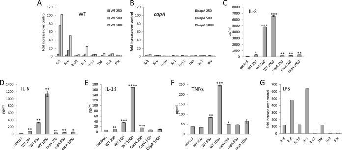
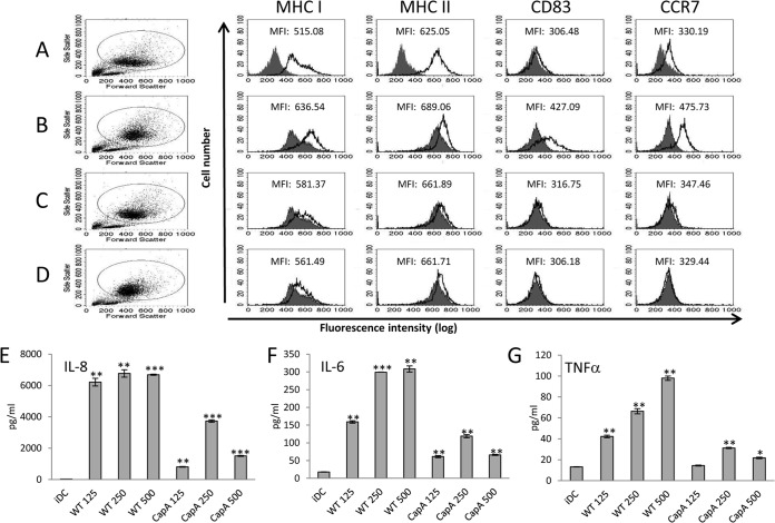
The antiphagocytic capsule of Bacillus anthracis is a major virulence factor. We hypothesized that it may also mediate virulence through inhibition of the host's immune responses. During an infection, the capsule exists attached to the bacterial surface but also free in the host tissues. We sought to examine the impact of free capsule by assessing its effects on human monocytes and immature dendritic cells (iDCs). Human monocytes were differentiated into iDCs by interleukin-4 (IL-4) and granulocyte-macrophage colony-stimulating factor (GM-CSF) over 7 days in the presence of capsule derived from wild-type encapsulated B. anthracis Ames (WT) or a control preparation from an isogenic B. anthracis Ames strain that produces only 2% of the capsule of the WT (capA mutant). WT capsule consistently induced release of IL-8 and IL-6 while the capA mutant control preparation elicited either no response or only a minimal release of IL-8. iDCs that were differentiated in the presence of WT capsule had increased side scatter (SSC), a measure of cellular complexity, when assessed by flow cytometry. iDCs differentiated in the presence of WT capsule also matured less well in response to subsequent B. anthracis peptidoglycan (Ba PGN) exposure, with reduced upregulation of the chemokine receptor CCR7, reduced CCR7-dependent chemotaxis, and reduced release of certain cytokines. Exposure of naive differentiated control iDCs to WT capsule did not alter cell surface marker expression but did elicit IL-8. These results indicate that free capsule may contribute to the pathogenesis of anthrax by suppressing the responses of immune cells and interfering with the maturation of iDCs.


Figures

FIG 1
FIG 1
Capsule expression is greatly reduced in the capA mutant B. anthracis strain. Capsule was purified from broth culture supernatants as described in Materials and Methods. Fifty micrograms of capsule from WT (lane 1) and capA mutant (lane 2) per lane was electrophoresed on a 1% agarose gel, and the gel was stained with methylene blue.
FIG 2
FIG 2
B. anthracis capsule induces cytokine responses from differentiating human monocytes. (A to F) Differentiating human monocytes were exposed to 250 to 1,000 μg/ml of WT capsule (A, C, D, E, and F) or an equivalent amount of capA mutant preparation (B, C, D, E, and F) for 7 days. Medium samples collected on day 3 were analyzed for cytokines as described in Materials and Methods. (A and B) Data are presented as the fold increase over the basal cytokine levels released by unstimulated control cells cultured in parallel. Data for IL-8 (C), IL-6 (D), IL-1β (E), and TNF-α (F) from day 3 are also presented as mean pg/ml ± SEM (n = 2). Statistical significance of the IL-8, IL-6, IL-1β, and TNF-α responses compared to the unstimulated control was determined by t test (*, P < 0.05; **, P < 0.01; ***, P < 0.001; ****, P < 0.0001). (G) The comparable fold increase in cytokine response of differentiating human monocytes exposed to 10 ng/ml LPS is shown after 3 days of culture. Data from a representative donor are shown.
FIG 3
FIG 3
Exposure of monocytes to B. anthracis capsule during differentiation alters the phenotype of the resulting iDCs. Differentiating human monocytes were exposed to 125 to 500 μg/ml WT capsule or equivalent amounts of capA mutant preparation for 7 days. On day 7, the cells were assessed for MHC I, MHC II, CD83, CCR7, and SSC by flow cytometry. (A) The filled histograms represent the isotype control staining, and the black-outlined histograms represent the marker staining for the naive control iDCs. (B and C) The filled histograms represent the marker staining for the naive control iDCs. The black-outlined histograms represent the marker staining for the cells differentiated in the presence of 500 μg/ml WT capsule (B) or capA mutant preparation (C). P values were determined by comparing the means of the MFIs for the WT capsule- and capA mutant-treated cells for all donors tested to the mean MFI for the naive control iDCs (P > 0.05 for all markers, n = 6 donors). (D) The filled histograms represent the SSC for the naive control iDCs. The black-outlined histograms represent the SSC for the cells differentiated in the presence of WT capsule. Statistical significance of changes in SSC compared to the naive control was determined by t test (P < 0.01, for 5 of 6 donors). Data from a single donor are shown.
FIG 4
FIG 4
Differentiation in the presence of B. anthracis capsule results in DCs with reduced CCR7 expression and reduced CCR7-dependent chemotaxis upon maturation with Ba PGN. Human monocytes were differentiated into iDCs in the presence of 500 μg/ml WT capsule or an equivalent amount of capA mutant preparation for 7 days. Control cells were differentiated in parallel in the absence of capsule. On day 7, the cells were stimulated with 1 μg/ml Ba PGN to induce the mDC phenotype. Additional control cells were maintained in differentiation medium alone to preserve the iDC phenotype. On day 9, half of the cells were assessed for MHC I, MHC II, CD83, and CCR7 expression by flow cytometry (A, B, C, and D). (A) The filled histograms represent the isotype control staining, and the black-outlined histograms represent the marker staining for the naive control iDCs. (B) The filled histograms represent the unstimulated naive control iDCs. The black-outlined histograms represent control iDCs after stimulation with 1 μg/ml Ba PGN to induce the mDC phenotype. (C and D) The filled histograms represent the mDC control. The black-outlined histograms represent Ba PGN-stimulated cells that were exposed to 500 μg/ml WT capsule (C) or to capA mutant preparation (D). P values were determined by comparing the means of the MFI for the WT capsule- and capA mutant-treated cells for all donors tested to the mean MFI for the Ba PGN-matured controls (P < 0.05 only for CCR7 for WT capsule, n = 5 donors). Histograms and MFIs shown are from a single donor. (E and F) The other half of the cells was subjected to a migration assay using CCL19 (E) or CCL21 (F) as the chemoattractive ligand, and the chemotactic index was calculated as described in Materials and Methods. Data shown are the means of the chemotactic indices for all the donors tested (n = 4). Statistical significance was determined by t test (*, P < 0.05, n = 4 donors).
FIG 5
FIG 5
Exposure of monocytes to B. anthracis capsule during differentiation alters the resulting iDCs' cytokine response to Ba PGN. Human monocytes were differentiated into iDCs in the presence of 500 μg/ml capsule or capA mutant preparation for 7 days. Control cells were differentiated in parallel in the absence of capsule. On day 7, the treated and naive control cells were stimulated with 1 μg/ml Ba PGN. Additional control cells were maintained unstimulated. On day 9, the media were analyzed for cytokines. Several donors exhibited decreased release of IL-6 (A), IL-10 (B), IL-1β (C), TNF-α (D), IFN-γ (E), and IL-23 (F) upon stimulation with Ba PGN (*, P < 0.05; **, P < 0.01; ***, P < 0.001; n = 2). P values for the responses to the two preparations (WT and capA mutant) were determined by comparing the mean pg/ml of each cytokine detected for the treated cells to the mean pg/ml of each cytokine detected for the control cells for each of four or five donors in a two-tailed t test. Data shown are from a representative donor.
FIG 6
FIG 6
Exposure of naive iDCs to B. anthracis capsule does not induce an mDC phenotype but does elicit cytokines. Human monocytes were differentiated into iDCs over the course of 7 days. On day 7, the iDCs were exposed to 500 μg/ml WT capsule or an equivalent amount of capA mutant preparation. On day 9, the cells were assessed for MHC I, MHC II, CD83, and CCR7 expression by flow cytometry (A, B, C, and D) and the media were analyzed for cytokines (E, F, and G). (A) The filled histograms represent the isotype control staining, and the black-outlined histograms represent the marker staining for the untreated naive control iDCs. (B) The filled histograms represent the unstimulated naive control iDCs, and the black-outlined histograms represent control iDCs that were stimulated with 100 ng/ml LPS to induce the mDC phenotype. (C and D) The filled histograms represent the unstimulated naive control iDCs. The black-outlined histograms represent cells that were exposed to 500 μg/ml WT capsule (C) or to capA mutant preparation (D). P values were determined by comparing the means of the MFI for the WT capsule and capA mutant-treated cells for all donors tested to the mean MFI for the unstimulated naive control iDCs (P > 0.05 for all markers, n = 3 donors). (E, F, and G) Data are presented as mean pg/ml ± SEM (n = 2) for IL-8 (E), IL-6 (F), and TNF-α (G). Statistical significance of the IL-8, IL-6, and TNF-α responses compared to the unstimulated iDC control was determined by t test (*, P < 0.05; **, P < 0.01; ***, P < 0.001). Data from a single donor are shown.

Similar articles

Cited by

References

    1. Mikesell P, Ivins BE, Ristroph JD, Dreier TM. 1983. Evidence for plasmid-mediated toxin production in Bacillus anthracis. Infect. Immun. 39:371–376. - PMC - PubMed
    1. Green BD, Battisti L, Koehler TM, Thorne CB, Ivins BE. 1985. Demonstration of a capsule plasmid in Bacillus anthracis. Infect. Immun. 49:291–297. - PMC - PubMed
    1. Uchida I, Sekizaki T, Hashimoto K, Terakado N. 1985. Association of the encapsulation of Bacillus anthracis with a 60 megadalton plasmid. J. Gen. Microbiol. 131:363–367. - PubMed
    1. Makino S, Uchida I, Terakado N, Sasakawa C, Yoshikawa M. 1989. Molecular characterization and protein analysis of the cap region, which is essential for encapsulation in Bacillus anthracis. J. Bacteriol. 171:722–730. - PMC - PubMed
    1. Scorpio A, Chabot DJ, Day WA, O'Brien DK, Vietri NJ, Itoh Y, Mohamadzadeh M, Friedlander AM. 2007. Poly-γ-glutamate capsule-degrading enzyme treatment enhances phagocytosis and killing of encapsulated Bacillus anthracis. Antimicrob. Agents Chemother. 51:215–222. 10.1128/AAC.00706-06. - DOI - PMC - PubMed

Publication types

LinkOut - more resources