. 2014 Nov;111(11):2239-51.
doi: 10.1002/bit.25282. Epub 2014 Jul 4.

Fractional factorial design to investigate stromal cell regulation of macrophage plasticity

Affiliations

Fractional factorial design to investigate stromal cell regulation of macrophage plasticity

Jeffrey A Barminko et al. Biotechnol Bioeng. 2014 Nov.

Abstract

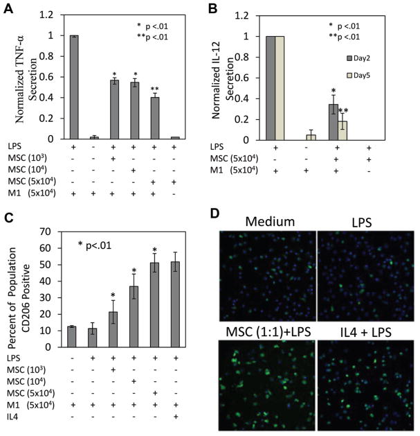
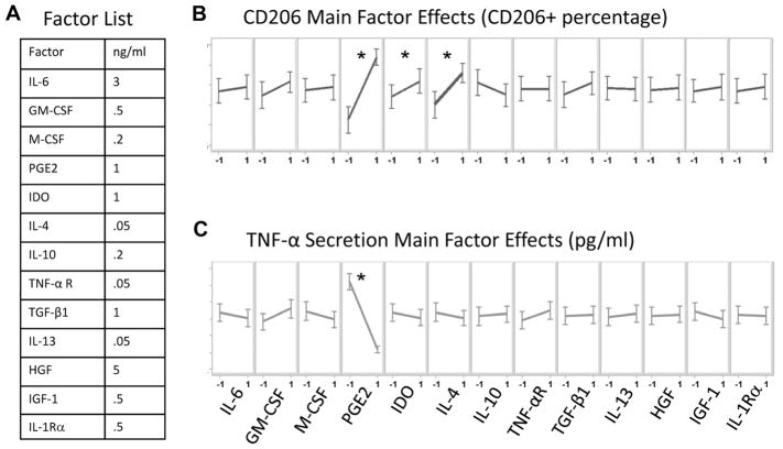
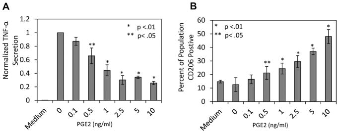
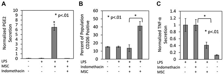
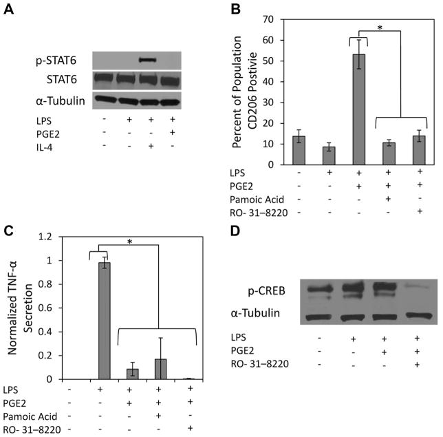
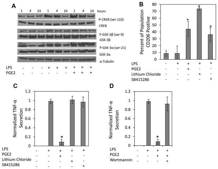
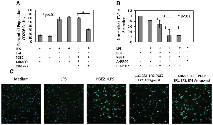
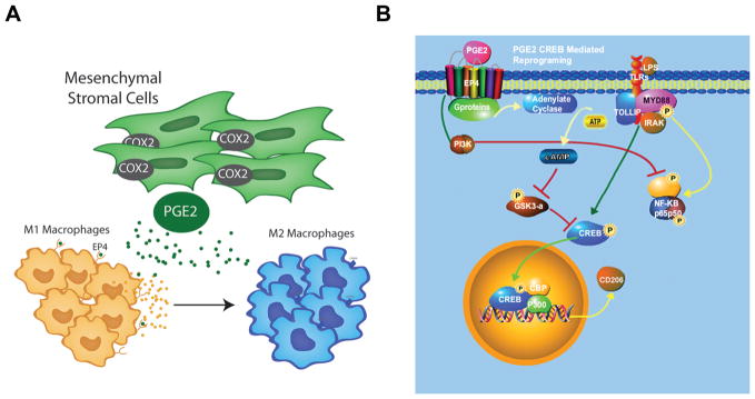
Understanding the regulatory networks which control specific macrophage phenotypes is essential in identifying novel targets to correct macrophage mediated clinical disorders, often accompanied by inflammatory events. Since mesenchymal stromal cells (MSCs) have been shown to play key roles in regulating immune functions predominantly via a large number of secreted products, we used a fractional factorial approach to streamline experimental evaluation of MSC mediated inflammatory macrophage regulation. Our macrophage reprogramming metrics, human bone marrow MSC attenuation of macrophage pro-inflammatory M1 TNFα secretion and simultaneous enhanced expression of the M2 macrophage marker, CD206, were used as analysis endpoints. Objective evaluation of a panel of MSC secreted mediators indicated that PGE2 alone was sufficient in facilitating macrophage reprogramming, while IL4 only provided partial reprogramming. Inhibiting stromal cell PGE2 secretion with Indomethacin, reversed the macrophage reprogramming effect. PGE2 reprogramming was mediated through the EP4 receptor and indirectly through the CREB signaling pathway as GSK3 specific inhibitors induced M1 macrophages to express CD206. This reprogramming pathway functioned independently from the M1 suppression pathway, as neither CREB nor GSK3 inhibition reversed PGE2 TNF-α secretion attenuation. In conclusion, fractional factorial experimental design identified stromal derived PGE2 as the factor most important in facilitating macrophage reprogramming, albeit via two unique pathways.

Keywords: PGE2; fractional factorial design; macrophage; mesenchymal stromal cells.


Figures

Figure 1
Figure 1
MSC attenuate macrophage pro-inflammatory cytokine secretion. A: At day 2 M1 macrophage TNF-α secretion was diminished with MSC co-culture. There was a concentration dependent reduction in TNF-α, maximum attenuation (55% reduction) was achieved at a: MSC/M1 ratio of 1:1. *Represents statistical significance relative to LPS and ** to MSC/M1 ratios of 1:50 and 1:5. B: MSCs attenuate M1 macrophage IL12 (p40) secretion at days 2 and 5 after LPS stimulation. *Represents statistical significance relative to LPS and ** to MSC/M1 day 2 cultures. C: MSC promote M1 macrophages stimulated with LPS to express CD206. At day 2 MSC co-culture resulted in a significant increase in the percentage of macrophages expressing CD206 relative to LPS and medium conditions. There was a dose dependent increase in the percentage of CD206 expressing cells which plateaued at ~50%. IL4 (10 ng/mL) promoted CD206 expression to approximately the same level as PGE2. *,**Represent statistical significance relative to LPS. D: Images depicting the elevated levels of CD206 observed when M1 macrophages are treated with MSC and IL4.
Figure 2
Figure 2
Fractional factorial evaluation of MSC factor effects on M1 function. A: List of MSC secreted factors and their concentrations evaluated in the study. A 128-condition experiment was designed to identify factors which may mediate MSC regulation of M1 macrophages. Conditions were made, spiked with LPS and transferred to M1 macrophage 96 well cultures. After 48 h cells were immunostained for CD206 and TNF-α levels were measured via ELISA. B and C: Illustrate main factor effects on the metrics CD206 and TNF-α, respectively. −1 and 1 designate the absence or presence of the factor, respectively. The slope of the line conveys the magnitude of change in the presence of a particular factor. Differences were considered statistically significant based on a confidence interval of 95% (P < 0.05). PGE2 and IL4 demonstrated the ability to significantly promote CD206. However, only PGE2 was able to significantly attenuate TNF-α secretion. *P < 0.05 represent statistical significant based on one-way ANOVA
Figure 3
Figure 3
PGE2 attenuates M1 TNF-α secretion and promotes M1 CD206 expression. M1 macrophages were stimulated with LPS and immediately treated with increasing concentrations of PGE2, except for the none treated control labeled Medium. At day 2 macrophage (A) TNF-α secretion and (B) CD206 expression were quantified. Increasing concentration of PGE2 attenuated M1 macrophage TNF-α secretion, which plateaued at 2.5 ng/mL at a reduction of ~75%. The percentage of CD206 expressing macrophages displayed the reverse relationship to increasing PGE2 concentration, however displayed a more linear response, and reached a maximum value of ~50%. *,**Represent statistical significance of P < 0.01 and P < 0.05 relative to LPS conditions respectively.
Figure 4
Figure 4
MSC PGE2 secretion promotes macrophage reprogramming. A: Supernatant from M1: MSC co-cultures treated with LPS (1 μg/mL) were assessed for PGE2 concentration. M1 macrophages in basal and stimulated conditions did not produce PGE2. However, in the presence of MSC ~6.5 ng/mL was detected. When a COX2 inhibitor, indomethacin (10 μM), is administered to the co-culture MSC PGE2 secretion is eliminated. *Represents statistical significance (P < 0.01) relative to LPS, medium and indomethacin. B: CD206 expression and (C) TNF-α secretion were assessed with indomethacin present in the MSC:M1 co-culture. The inhibition of MSC PGE2 secretion prevented the percent increase of CD206 expressing cells as well as the attenuation of TNF-α. *Represents statistical significance (P < 0.01) between MSC:M1 conditions in the presence and absence of indomethacin.
Figure 5
Figure 5
PGE2 promotes increased M1 macrophage CD206 expression through CREB signaling. A: M1 macrophages were stimulated with LPS (1 μg/mL) and treated with IL-4 (10 ng/mL) or PGE2 (10 ng/mL) for 15 min before protein extraction. Immunoblotting for phosphorylated STAT6 at tyr-641 showed that PGE2, unlike IL4, does not promote STAT6 activation. B: M1 macrophages were pre-incubated with RO-31-8220 for 10 min, before stimulation and treatment with PGE2 (10 ng/mL). Pamoic acid was administered at the time of LPS stimulation and PGE2 treatment. Inhibition of CREB signaling with Pamoic acid or RO-31-8220 prevented MSC promotion of M1 CD206 expression, but had no effect on (C) TNF-α secretion. D: Immunoblotting for phosphorylated CREB, at ser-133 15 min after LPS stimulation and treatment with PGE2 did not show increased expression of pCREB. LPS stimulation alone was able to significantly increase p-CREB expression. Immunoblotting confirmed RO-31-8220 inhibition of CREB activation.
Figure 6
Figure 6
PGE2 inhibits GSK-3α and leads to increased M1 CD206 expression. A: Protein samples from M1 macrophages stimulated with LPS and treated with PGE2 (10 ng/mL) for 1, 4, and 24 h were immunoblotted for GSK, CREB, and STAT6 phosphorylation. PGE2 treatment increased the expression of phosphorylated GSK-3α at ser-21 over 24 h. No significant differences were observed in the expression of phosphorylated GSK-3β at ser-9 in any condition. PGE2 treatment modestly prevented CREB de-phosphorylation overtime. B and C: M1 macrophages were pre-treated with GSK inhibitors, lithium chloride (40 mM) and SB415286 (40 μM), for 1 h before LPS stimulation. Two days of LPS stimulation occurred in the presence of the inhibitors and led to an increase in the percentage of (B) CD206 positive cells in the M1 macrophage population, but had no effect on (C) TNF-α Secretion. D: M1 macrophage were stimulated with LPS and treated with PGE2 in the presence of a PI3k inhibitor, which reversed PGE2 attenuation of M1 macrophage TNF-α secretion. *Represents statistical significance (P < 0.01) relative to LPS conditions.
Figure 7
Figure 7
PGE2 mediates M1 macrophage reprogramming through the EP4 receptor. LPS stimulated M1 macrophages were treated with IL4 (10 ng/mL), PGE2 (10 ng/mL), PGE2 with an EP4 antagonist (L161982) or a general antagonist to the other EP receptors (AH6809). A: The increase in the percentage of CD206 positive cells previously observed with PGE2 treatment was diminished with an EP4 antagonist. Antagonists to the other EP receptors did not interfere with PGE2 promotion of CD206. B: PGE2 attenuation of TNF-α was also reversed with an EP4 antagonist. Blocking the other EP receptors did not reverse PGE2 TNF-α attenuation. C: Representative images of CD206 depicting the reduction of percent CD206 positive cells when the EP4 receptors is blocked during PGE2 treatment.
Figure 8
Figure 8
MSC PGE2 secretion reprograms M1 via two independent pathways to M2 macrophages. A: MSCs secrete PGE2, which binds to the EP4 receptors on M1 macrophages and facilitates their transition to M2 macrophages. B: PGE2 binding to EP4 receptors activates two independent pathways (1) the PI3k pathway which inhibits NF-kβ simulation of pro-inflammatory cytokines such as TNF-α and IL12 and (2) the cAMP pathway which leads to the inhibition of GSK and subsequent activation of CREB signaling, which leads to the transcription of genes associated with the M2 macrophage phenotype.

Similar articles

Cited by

References

    1. Best JL, Amezcua CA, Mayr B, Flechner L, Murawsky CM, Emerson B, Zor T, Gardner KH, Montminy M. Identification of small-molecule antagonists that inhibit an activator: Coactivator interaction. Proc Natl Acad Sci USA. 2004;101:17622–17627. - PMC - PubMed
    1. Beurel E, Michalek SM, Jope RS. Innate and adaptive immune responses regulated by glycogen synthase kinase-3 (GSK3) Trends Immunol. 2010;31:24–31. - PMC - PubMed
    1. Bouffi C, Bony C, Courties G, Jorgensen C, Noel D. IL-6-dependent PGE2 secretion by mesenchymal stem cells inhibits local inflammation in experimental arthritis. PLoS ONE. 2010;5:e14247. - PMC - PubMed
    1. Bullock BP, Habener JF. Phosphorylation of the cAMP response element binding protein CREB by cAMP-dependent protein kinase A and glycogen synthase kinase-3 alters DNA-binding affinity, conformation, and increases net charge. Biochemistry. 1998;37:3795–3809. - PubMed
    1. Chen Y, Bloemen V, Impens S, Moesen M, Luyten FP, Schrooten J. Characterization and optimization of cell seeding in scaffolds by factorial design: Quality by design approach for skeletal tissue engineering. Tissue Eng Part C Methods. 2011;17:1211–1221. - PubMed

Publication types

MeSH terms

LinkOut - more resources