Review
. 2014 Sep;1323(1):115-39.
doi: 10.1111/nyas.12462. Epub 2014 May 30.

Continuing challenges in influenza

Affiliations
Review

Continuing challenges in influenza

Robert G Webster et al. Ann N Y Acad Sci. 2014 Sep.

Abstract

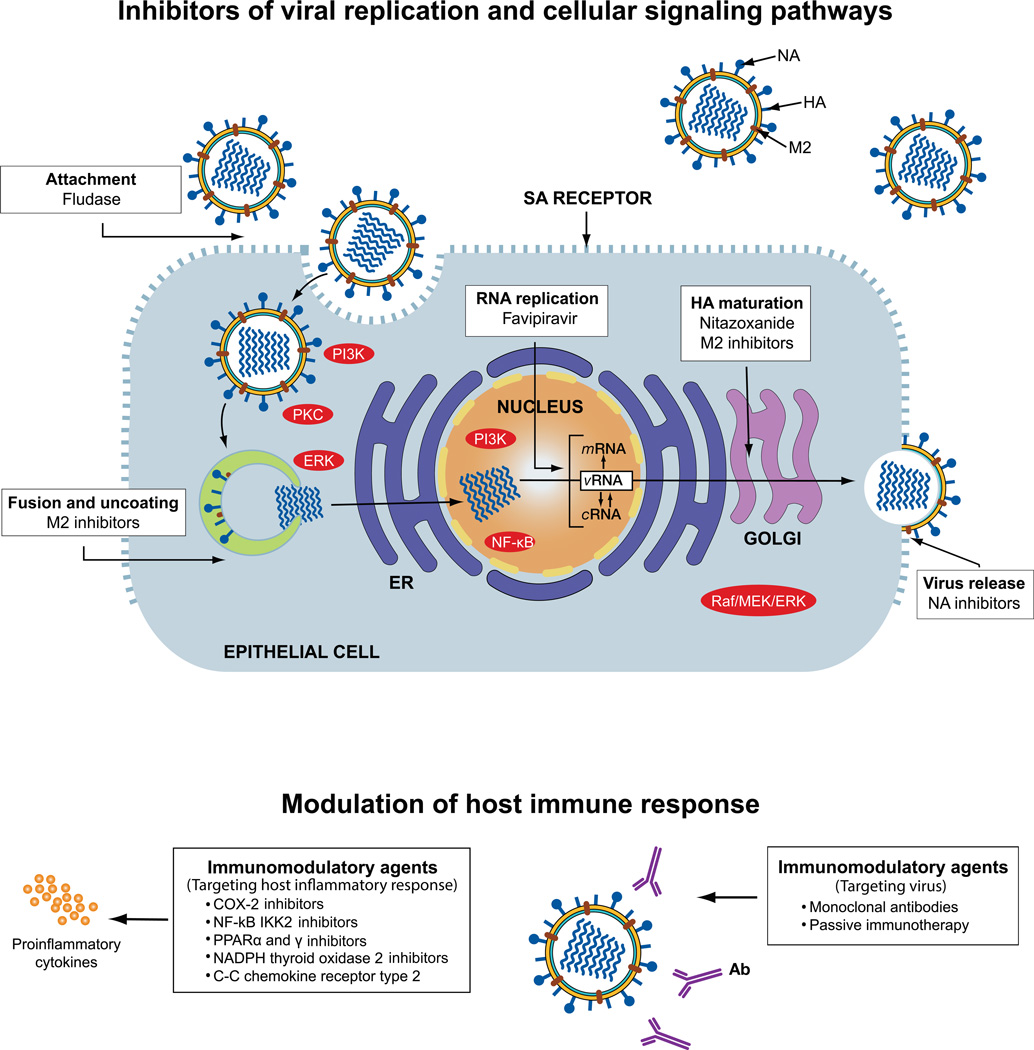
Influenza is an acute respiratory disease in mammals and domestic poultry that emerges from zoonotic reservoirs in aquatic birds and bats. Although influenza viruses are among the most intensively studied pathogens, existing control options require further improvement. Influenza vaccines must be regularly updated because of continuous antigenic drift and sporadic antigenic shifts in the viral surface glycoproteins. Currently, influenza therapeutics are limited to neuraminidase inhibitors; novel drugs and vaccine approaches are therefore urgently needed. Advances in vaccinology and structural analysis have revealed common antigenic epitopes on hemagglutinins across all influenza viruses and suggest that a universal influenza vaccine is possible. In addition, various immunomodulatory agents and signaling pathway inhibitors are undergoing preclinical development. Continuing challenges in influenza include the emergence of pandemic H1N1 influenza in 2009, human infections with avian H7N9 influenza in 2013, and sporadic human cases of highly pathogenic avian H5N1 influenza. Here, we review the challenges facing influenza scientists and veterinary and human public health officials; we also discuss the exciting possibility of achieving the ultimate goal of controlling influenza's ability to change its antigenicity.

Keywords: H1N1; H5N1; H7N9; antigenic changes; antiviral drugs; influenza virus; pandemic; vaccines; zoonosis.


Figures

Figure 1
Figure 1
A diagram of the host ranges of the 18 hemagglutinin (HA) subtypes of influenza A viruses. Open figures indicate hosts in which stable lineages are established. Filled figures indicate hosts in which sporadic but not consistent interhost transmission of influenza has occurred. Colored figures indicate influenza subtypes that have caused pandemic or epidemic influenza. The 16 HA subtypes of influenza A viruses in aquatic birds and the two subtypes in bats cause inapparent disease and are shed in feces. In mammalian hosts, influenza causes upper respiratory disease. In horses, two subtypes—Equine 1 (H7N7) and Equine-2 (H3N8)—have circulated; however, Equine-1 has not been detected in horses for the past 2 decades. Sporadic transmission of H2 in pigs in China was reported in 1957, when the Asian H2N2 pandemic occurred but did not establish a stable lineage. Sporadic transmissions of H4, H5, H6, H7, H9, and H10 to humans and pigs have increased since the mid-1990s, but no consistent interhost transmission has occurred.
Figure 2
Figure 2
Influenza pandemics in humans and pigs in the past century. Only three subtypes of influenza A viruses have caused pandemics in humans: H1N1, H2N2, and H3N2. H1N1 and H3N2 have also caused outbreaks in swine during the same period. On the basis of seroarcheological studies, it is proposed that H2N2 and H3N2 subtypes have previously infected humans. The most severe pandemic, which was caused by the Spanish influenza of 1918, resulted in more than 40 million deaths worldwide, many of which resulted from co-infection with bacteria. Subsequent pandemics have been less severe. Because H2N2 viruses have not circulated in humans for nearly 50 years, most people are immunologically naive. The H2N2 viruses, however, continue to circulate in aquatic birds. The jagged edges on the ends of the H1N1pdm09 and H3N2 bars indicate that these viruses continue to circulate.
Figure 3
Figure 3
The hemagglutinin (HA) molecule of A/Aichi/2/68 (H3N2) influenza virus in which the amino acid residues that are crucial in antigenic drift are indicated. (A) The trimeric model of the HA molecule. One monomer is shown in black; key amino acids in antigenic drift are shown in red; and the receptor-binding site is shown in yellow. (B) Residue 145 is located in antigenic site A, and residues 155, 156, 158, 159, and 193 are in antigenic site B. These are considered “cluster-transition” amino acids and correspond with amino acids in the HA of H1N1 and influenza B viruses. An asterisk indicates accessory substitutions. Reprinted from Ref. with permission from the AAAS.
Figure 4
Figure 4
Monoclonal antibodies are cross-reactive with hemagglutinins (HAs) of influenza A viruses of groups 1 and 2. (A–B) Phylogenetic trees of the 16 HA influenza A virus subtypes classified into groups 1 and 2 are shown. Phylogenetic trees with HA subtypes bound or neutralized by S139/1 (A) or FI6 (B) are highlighted in red. Note that both antibodies neutralize subtypes from both major virus groups. (C) Like CR6261 and CR8020, FI6 also recognizes an epitope on the HA stem. HA is depicted as a molecular surface, with the HA1 and HA2 subunits from one protomer highlighted in pink and cyan, respectively. The heavy and light chains of FI6 are shown as red and yellow ribbons, respectively. Although the precise location of its epitope is unknown, escape mutations suggest that S139/1 recognizes the HA1 head region. (D) The epitopes of FI6 and CR6261/F10 overlap extensively (orange surface), and only a small number of HA residues are recognized by FI6 only (green surface) or CR6261 only (blue surface). Reprinted from Ref. with permission from Elsevier.
Figure 5
Figure 5
Schematic representation of influenza virus replication cycle and drugs currently used for prophylaxis and/or treatment of influenza, drugs in clinical trials, and those in preclinical development. Antiviral therapy predominately relies on virus-specific drugs such as the neuraminidase (NA) inhibitors (NAIs) oseltamivir and zanamivir, which bind the enzymatically active site of the NA of influenza A or B viruses and inhibit the release of budding viral particles from infected host cells. M2 inhibitors (amantadine and rimantadine) block the ion channel activity of the M2 protein of influenza A virus at the stage of virus fusion and uncoating. Drugs undergoing clinical trials in the U.S. for the prevention and treatment of influenza include Fludase (DAS181), Favipiravir (T-705), and Nitazoxanide. Sialidase inhibitor Fludase cleaves sialic acid (SA) α2,3- and SA α2,6-linked cellular receptors and inhibits virus attachment to them. Prodrug favipiravir is converted intracellularly to its active metabolite, T-705 ribofuranosyltriphosphate, which inhibits RNA polymerase activity. Nitazoxanide affects the terminal glycosylation of the hemaggluttinin (HA) glycoprotein, thereby impairing HA trafficking between the endoplasmic reticulum (ER) and the Golgi complex. The host’s immune response can be modulated by inhibitors of various cellular signaling pathways and cascades during the virus’ replication cycle. Although several inhibitors are currently in preclinical development, these investigational immunomodulatory agents can be categorized into two groups based on their target: those that target the host’s inflammatory response (left) and those that target the virus (right). Ab, antibody; COX2, cyclooxygenase-2; cRNA, complementary RNA; ERK, extracellular signaling–regulated kinase; HA, hemagglutinin; NA, neuraminidase; M2, matrix protein; MEK, MAP/ERK kinase; mRNA, viral messenger RNA; PI3K, phosphatidylinositol-3 kinase; PPAR, peroxisome proliferator-activated receptor; PKC, protein kinase C; vRNA, viral RNA.

Similar articles

Cited by

References

    1. Shaw ML, Palese P. Orthomyxoviradae: the viruses and their replication. In: Knipe D, Howley P, editors. Fields’ Virology, Sixth Edition. Philadelphia, PA: Lippin Williams & Wilkins; 2011. pp. 1647–1689.
    1. Webster RG, Bean WJ, Gorman OT, et al. Evolution and ecology of influenza A viruses. Microbiol. Rev. 1994;56:152–179. - PMC - PubMed
    1. Jernigan DB, Cox NJ. Human influenza: One Health, One World. In: Webster RG, Monto AS, Braciale TJ, Lamb RA, editors. Textbook of Influenza, Second Edition. West Sussex: John Wiley and Sons; 2013. pp. 3–19.
    1. Russell RJ, Kerry PS, Stevens DJ, et al. Structure of influenza hemagglutinin in complex with an inhibitor of membrane fusion. Proc. Natl. Acad. Sci. U.S.A. 2008;105:17736–17741. - PMC - PubMed
    1. Tong S, Zhu X, Li Y, et al. New world bats harbor diverse influenza A viruses. PLoS Pathog. 2013;9:e1003657. - PMC - PubMed

Publication types