. 2014 Aug;196(15):2889-900.
doi: 10.1128/JB.01516-14. Epub 2014 Jun 2.

Localization of the outer membrane protein OmpA2 in Caulobacter crescentus depends on the position of the gene in the chromosome

Affiliations

Localization of the outer membrane protein OmpA2 in Caulobacter crescentus depends on the position of the gene in the chromosome

Luis David Ginez et al. J Bacteriol. 2014 Aug.

Abstract

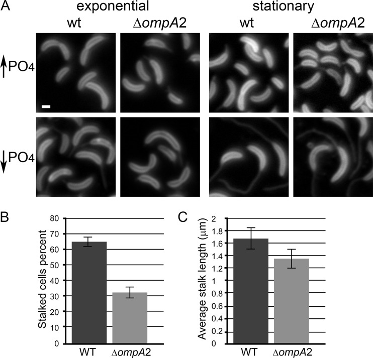
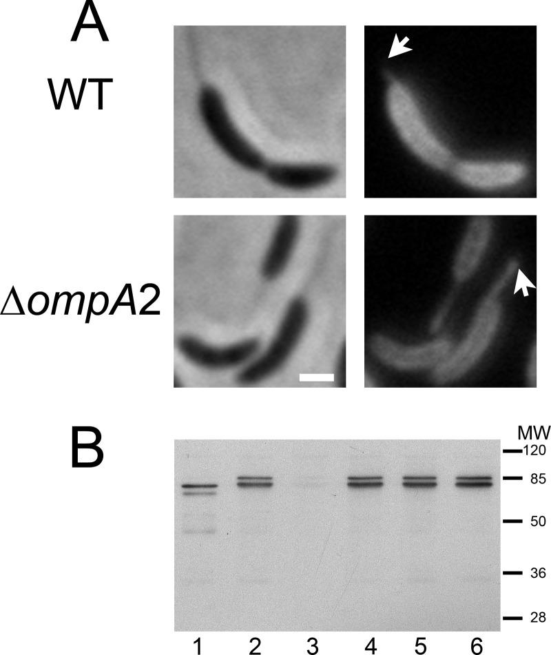
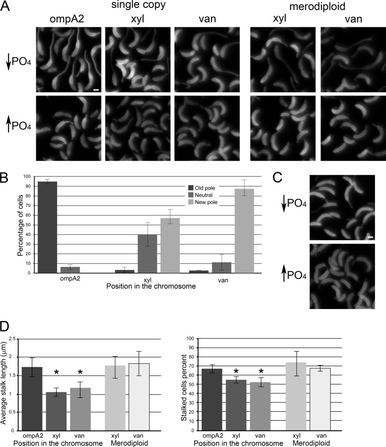
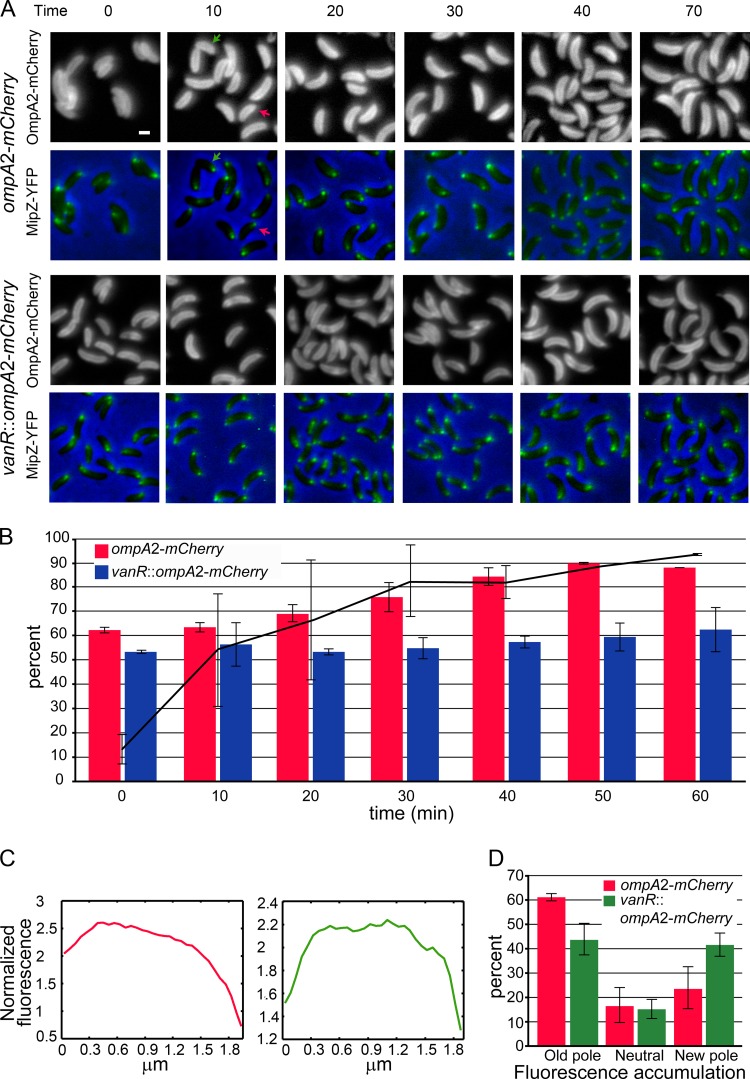
The outer membrane of Gram-negative bacteria is an essential structure involved in nutrient uptake, protection against harmful substances, and cell growth. Different proteins keep the outer membrane from blebbing out by simultaneously interacting with it and with the cell wall. These proteins have been mainly studied in enterobacteria, where OmpA and the Braun and Pal lipoproteins stabilize the outer membrane. Some degree of functional redundancy exists between these proteins, since none of them is essential but the absence of two of them results in a severe phenotype. Caulobacter crescentus has a different strategy to maintain its outer membrane, since it lacks the Braun lipoprotein and Pal is essential. In this work, we characterized OmpA2, an OmpA-like protein, in this bacterium. Our results showed that this protein is required for normal stalk growth and that it plays a minor role in the stability of the outer membrane. An OmpA2 fluorescent fusion protein showed that the concentration of this protein decreases from the stalk to the new pole. This localization pattern is important for its function, and it depends on the position of the gene locus in the chromosome and, as a consequence, in the cell. This result suggests that little diffusion occurs from the moment that the gene is transcribed until the mature protein attaches to the cell wall in the periplasm. This mechanism reveals the integration of different levels of information from protein function down to genome arrangement that allows the cell to self-organize.


Figures

FIG 1
FIG 1
The ompA2 mutant has an unstable OM. The growth rate, sensitivity to SDC, and outer membrane stability of the ompA2 mutant were determined. (A) Growth rates (expressed as generation time) of the wild-type (WT) and ompA2 mutant strains under different growth conditions. Error bars represent standard deviations, and significantly different growth rates are indicated with asterisks. (B) Growth curve for the wild-type and ompA2 mutant strains in the presence of SDC (SDC was present from the beginning of the experiment). (C) Wild-type and ompA2 mutant cells stained with FM 4-64FX. The arrow indicates a small vesicle. White bar, 1 μm.
FIG 2
FIG 2
OmpA2 is required for normal stalk growth. The average stalk length and number of prosthecated cells were determined for the wild-type (WT) and ompA2 mutant strains. (A) Wild-type and ompA2 mutant cells from exponentially growing (OD660 of 0.3) and stationary cultures were stained with FM 4-64FX. White bar, 1 μm. (B) The number of stalked cells from exponentially growing cultures in low-phosphate medium. (C) The average lengths of the stalks of exponentially growing cells in low-phosphate medium. Error bars are based on results from three independent experiments, in each of which 150 cells were analyzed.
FIG 3
FIG 3
OmpA2 is an OM protein that forms a concentration gradient. The localization pattern of the OmpA2-mCherrry protein fusion was determined, and its insertion in the OM was verified. (A) Bright-field and fluorescent images of cells expressing OmpA2-mCherry from its native promoter. Cells were taken from exponentially growing cultures in high-phosphate or low-phosphate medium. (B) Plasmolysis of cells expressing OmpA2-mCherry and the IM protein TolQ fused to YFP (red and green, respectively). The top and bottom panels show cells before and after plasmolysis, respectively. White bar, 1 μm.
FIG 4
FIG 4
OmpA2-mCherry is evenly distributed when it is expressed from a plasmid. The localization pattern of OmpA2-mCherry expressed from a replicating plasmid is shown, along with Western blotting results for OmpA2-mCherry expressed from different loci and a plasmid. (A) Bright-field and fluorescent images of wild-type and ompA2 mutant cells expressing OmpA2-mCherry from a plasmid. White arrows indicate stalks partially labeled by the OmpA2-mCherry protein. White bar, 1 μm. (B) Western blot results for OmpA2-mCherry expressed from different chromosomal loci or a plasmid. Lanes: 1, CB15N/pRVompA2mCh-6; 2, CB15N ompA2::pompA2-mCh; 3, CB15N; 4, CB15N ompA2::pompA2-mCh; 5, CB15N xylR::pXpompA2-mCh; 6, CB15N vanR::pVpompA2-mCh.
FIG 5
FIG 5
The localization and function of OmpA2 depend on the position of the gene locus. The position of the ompA2-mCherry gene was changed to different loci and the location and function of the protein in stalk growth were evaluated. (A) Localization pattern of OmpA2-mCherry expressed from its native (ompA), xylR (xyl), or vanR (van) locus in single copy or as a second copy (merodiploid). Cells were grown in high- or low-phosphate medium and visualized when the culture reached an OD660 of 0.3. (B) The number of cells showing an accumulation at the old pole, at the new pole, or with an even distribution of OmpA2-mCherry expressed from different positions in the chromosome. (C) Localization pattern of another integral OM protein. The fluorescent images show cells expressing CC1750-mCherry from its native promoter. Cells were grown in high- or low-phosphate medium and visualized when the culture reached an OD660 of 0.3. (D) The average stalk length and percentage of stalked cells present in cultures of cells expressing OmpA2-mCherry from different loci. All quantifications were done with cells grown in low-phosphate medium, and images were taken when the culture reached an OD660 of 0.3. The bars represent the averages obtained from three independent experiments, in each of which at least 200 stalked predivisional cells (B) or 300 cells (D) were analyzed. The asterisks in panel D indicate averages significantly different from those obtained with cells expressing OmpA2-mChery from its native locus. Error bars represent standard deviations of three independent experiments. White bars (A and C), 1 μm.
FIG 6
FIG 6
The OmpA2 gradient forms before chromosome segregation. The localization of OmpA2-mCherry was followed during a cell cycle of a synchronized culture. (A) Independent time-lapse images of cells, grown in high-phosphate medium, that expressed MipZ-YFP and OmpA2-mCherry from its native or vanR locus. Green arrow, a cell with an even fluorescence distribution; red arrow, a cell with a fluorescence gradient. (B) Percentages of cells in a synchronized culture, demonstrating the fluorescence gradient. The black line indicates the percentages of cells with two MipZ-YFP foci. (C) The OmpA2-mCherry fluorescence intensity profile along the cell. The red and green profiles correspond to the cells marked by the red and green arrows in panel A. (D) Percentages of antibiotic-treated swarmer cells that showed a normal, inverse gradient or an even fluorescence distribution. The bars in all cases represent the averages obtained from three independent experiments in each of which at least 150 cells were analyzed. Error bars represent the standard deviations from three independent experiments.

Similar articles

Cited by

References

    1. Silhavy TJ, Kahne D, Walker S. 2010. The bacterial cell envelope. Cold Spring Harb. Perspect. Biol. 2:a000414. 10.1101/cshperspect.a000414 - DOI - PMC - PubMed
    1. Sonntag I, Schwarz H, Hirota Y, Henning U. 1978. Cell envelope and shape of Escherichia coli: multiple mutants missing the outer membrane lipoprotein and other major outer membrane proteins. J. Bacteriol. 136:280–285 - PMC - PubMed
    1. Braun V, Bosch V. 1972. Repetitive sequences in the murein-lipoprotein of the cell wall of Escherichia coli. Proc. Natl. Acad. Sci. U. S. A. 69:970–974. 10.1073/pnas.69.4.970 - DOI - PMC - PubMed
    1. Braun V, Rehn K. 1969. Chemical characterization, spatial distribution and function of a lipoprotein (murein-lipoprotein) of the E. coli cell wall. The specific effect of trypsin on the membrane structure. Eur. J. Biochem. 10:426–438 - PubMed
    1. Braun V, Wolff H. 1970. The murein-lipoprotein linkage in the cell wall of Escherichia coli. Eur. J. Biochem. 14:387–391. 10.1111/j.1432-1033.1970.tb00301.x - DOI - PubMed

Publication types

MeSH terms

Substances

LinkOut - more resources