

Case Reports
doi: 10.1212/WNL.0000000000000474.
FLAIR vascular hyperintensity resolution in a TIA patient: clinical-radiologic correlation
Affiliations
- PMID: 24891542
- PMCID: PMC4105261
- DOI: 10.1212/WNL.0000000000000474
Item in Clipboard
Case Reports
FLAIR vascular hyperintensity resolution in a TIA patient: clinical-radiologic correlation
Neurology.
.
Abstract
An 83-year-old woman presented with acute aphasia. Brain MRI, performed 3 hours after symptom onset, showed isolated fluid-attenuated inversion recovery vascular hyperintensities (FVH) in the left middle cerebral artery, including dot-like and serpentine hyperintensities (figure). Immediately after this first MRI (i.e., 3 hours and 15 minutes after symptom onset), aphasia resolved. A second MRI performed 15 minutes later showed FVH disappearance.
Figures
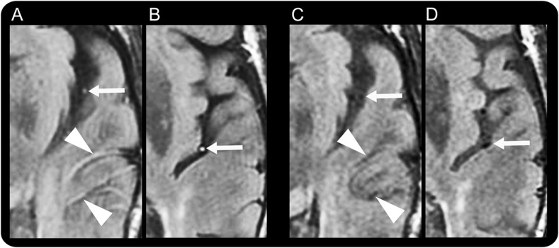
Fluid-attenuated inversion recovery vascular hyperintensities (FVH) during aphasia, including dot-like (arrows in A and B) and serpentine (arrowheads in A) hyperintensities, were seen in the middle cerebral artery branches. There were no abnormalities on diffusion-weighted or gradient echo images or magnetic resonance angiography. After aphasia resolution, the MRI showed FVH disappearance (C, D).
Comment in
-
FLAIR vascular hyperintensity resolution in a TIA patient: clinical-radiologic correlation.Neurology. 2015 Jan 13;84(2):215. doi: 10.1212/01.wnl.0000460112.49113.82. Neurology. 2015. PMID: 25583828 No abstract available.
-
Author response.Neurology. 2015 Jan 13;84(2):215. Neurology. 2015. PMID: 25729811 No abstract available.
References
-
- Kobayashi J, Uehara T, Toyoda K, et al. Clinical significance of fluid-attenuated inversion recovery vascular hyperintensities in transient ischemic attack. Stroke 2013;44:1635–1640 - PubMed
Publication types
MeSH terms
LinkOut - more resources
Full Text Sources
Other Literature Sources
Medical