. 2014 Aug;34(16):3106-19.
doi: 10.1128/MCB.00423-14. Epub 2014 Jun 2.

RNA-binding protein AUF1 promotes myogenesis by regulating MEF2C expression levels

Affiliations

RNA-binding protein AUF1 promotes myogenesis by regulating MEF2C expression levels

Amaresh C Panda et al. Mol Cell Biol. 2014 Aug.

Abstract

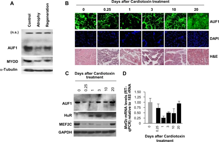
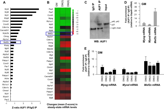
The mammalian RNA-binding protein AUF1 (AU-binding factor 1, also known as heterogeneous nuclear ribonucleoprotein D [hnRNP D]) binds to numerous mRNAs and influences their posttranscriptional fate. Given that many AUF1 target mRNAs encode muscle-specific factors, we investigated the function of AUF1 in skeletal muscle differentiation. In mouse C2C12 myocytes, where AUF1 levels rise at the onset of myogenesis and remain elevated throughout myocyte differentiation into myotubes, RNP immunoprecipitation (RIP) analysis indicated that AUF1 binds prominently to Mef2c (myocyte enhancer factor 2c) mRNA, which encodes the key myogenic transcription factor MEF2C. By performing mRNA half-life measurements and polysome distribution analysis, we found that AUF1 associated with the 3' untranslated region (UTR) of Mef2c mRNA and promoted MEF2C translation without affecting Mef2c mRNA stability. In addition, AUF1 promoted Mef2c gene transcription via a lesser-known role of AUF1 in transcriptional regulation. Importantly, lowering AUF1 delayed myogenesis, while ectopically restoring MEF2C expression levels partially rescued the impairment of myogenesis seen after reducing AUF1 levels. We propose that MEF2C is a key effector of the myogenesis program promoted by AUF1.


Figures

FIG 1
FIG 1
AUF1 levels during mouse skeletal muscle regeneration in vivo. (A) Western blot analysis of the levels of AUF1, MYOD, and loading control protein α-tubulin in mouse gastrocnemius muscle 14 days after suspension of the hind limbs (Atrophy) and 3 days after stopping the hind limb suspension to allow muscle regeneration (Regeneration). n.s., nonspecific. (B) Immunofluorescence detection of AUF1 in frozen muscle sections prepared at different time points during skeletal muscle regeneration in cardiotoxin-injured muscles. Sections were also stained with hematoxylin and eosin (H&E) and the DNA stain DAPI (4′,6-diamidino-2-phenylindole) to monitor the regeneration process. (C and D) Western blot analysis to check the expression of AUF1, HuR, and MEF2C (C) and RT-qPCR analysis to measure the levels of Mef2c mRNA (normalized to the levels of 18S rRNA) (D) during skeletal muscle regeneration in cardiotoxin-injured muscles (E). Data in panel D represent the means and standard errors of the means (SEM) from three different experiments.
FIG 2
FIG 2
C2C12 myoblast differentiation and expression of myogenic transcription factors. (A) Phase-contrast micrographs of undifferentiated (asynchronously growing) C2C12 myoblasts and C2C12 cells grown to confluence and differentiating into myotubes through culture in DMEM with 2% horse serum for up to 6 days. (B) Creatine kinase assay to quantify the degree of differentiation of C2C12 cells. (C) RT-qPCR analysis to monitor the expression of Myog, Myod, and Mef2c mRNAs, encoding myogenic factors, during C2C12 cell differentiation. The mRNA levels were normalized to the levels of 18S rRNA, measured by RT-qPCR analysis in the same samples. (D) Western blot analysis of the levels of AUF1 and myogenic transcription factors MYOD and MEF2C (as well as loading control GADPH) during differentiation of C2C12 myoblasts into myotubes. Data in panels B and C represent the means and SEM from three different experiments.
FIG 3
FIG 3
AUF1 interacts with mRNAs encoding skeletal muscle myogenesis factors. (A) AUF1 ribonucleoprotein immunoprecipitation to identify AUF1-bound mRNAs by microarray (RIP chip) analysis in C2C12 cells on DM(2); the levels of mRNAs in AUF1 IP were normalized to the levels of Gapdh mRNA and plotted as fold enrichment relative to the levels seen in control IgG IP samples. The graph shows the interaction of AUF1 with mRNAs encoding proteins involved in myogenesis. (B) Microarray analysis of the levels of mRNAs encoding muscle differentiation factors in C2C12 myoblasts (GM) and C2C12 cells at different stages during differentiation to myotubes [DM(3) and DM(6)]. Data are shown as mean Z-scores. (C) Western blot analysis of AUF1 in AUF1 IP samples used for RIP chip (A) and RIP followed by RT-qPCR analysis (D and E). (D and E) RIP followed by RT-qPCR analysis to assess AUF1-associated Myog, Myod, and Mef2c mRNAs in GM C2C12 myoblasts (D) and in differentiating [DM(0) through DM(6)] C2C12 cells (E). Data in panels D and E represent the means and SEM from three different experiments.
FIG 4
FIG 4
AUF1 regulates MEF2C expression by interacting with the Mef2c 3′UTR. (A) Biotinylated RNA fragments spanning the 5′UTR, coding region (CR), and 3′UTR of the Mef2c mRNA used for pulldown identify fragments with affinity for AUF1 using cytoplasmic C2C12 lysates; AUF1 was visualized by Western blotting. (B) Schematic of the dual-luciferase reporter plasmids psiCHECK2, the control vector expressing renilla luciferase (RL) and the internal control firefly luciferase (FL), and psiCHECK2-Mef2c(3′), the test vector bearing the Mef2c 3′UTR downstream of the RL coding region. (C) Influence of AUF1 on the expression of the reporter constructs shown in panel B. Forty-eight hours after transfecting C2C12 cells with AUF1 siRNA or Ctrl siRNA, AUF1 levels were reduced substantially (left), as detected by Western blotting. Twenty-four hours after the transfection of C2C12 cells with either AUF1 siRNA or Ctrl siRNA, each reporter plasmid was transfected, and the ratio of RL activity to FL activity was calculated 24 h after that. The relative RL/FL ratio of AUF1 siRNA-transfected cells relative to the RL/FL of Ctrl siRNA-transfected cells is indicated (right). Data represent the means and SEM from 3 independent experiments. Significance (P) is indicated.
FIG 5
FIG 5
AUF1 silencing reduces the size of Mef2c mRNA polysomes. (A and B) Forty-eight hours after siRNA transfection of C2C12 cells, polysomes in cytoplasmic extracts were fractionated through sucrose gradients (the arrow indicates the direction of sedimentation) (A), and the relative distribution of Gapdh mRNA, encoding a housekeeping protein, and Mef2c mRNA was measured by RT-qPCR analysis of RNA in gradient fractions and represented as the percentage of total RNA in the gradient (B). (C and D) Forty-eight hours after siRNA transfection, C2C12 cells were subjected to differentiation; at DM(6), polysomes were analyzed as explained for panels A and B. Data are representative of three independent experiments.
FIG 6
FIG 6
AUF1 enhances Mef2c gene transcription. (A and B) AUF1 was silenced as described for Fig. 4C; 48 h later [DM(0)] or at DM(6), the levels of AUF1, MEF2C, and loading control α-tubulin were studied by Western blotting (A) and the levels of Mef2 mRNA by RT-qPCR analysis (B). (C) AUF1 was overexpressed by using a pool of four plasmids (derived from pFlag-CMV2), each expressing one AUF1 isoform (see Materials and Methods); 48 h later, when C2C12 cells were in DM(0), the levels of AUF1, MEF2C, and loading control heat shock protein 90 (HSP90) were studied by Western blotting (left) and the levels of Mef2 mRNA by RT-qPCR analysis (right). (D) The stability of Mef2c mRNA (and stable control Gapdh mRNA) was assessed by incubating C2C12 cells prepared as described for panel A with actinomycin D to block de novo transcription, whereupon the Mef2c mRNA and Gapdh mRNA levels were measured by RT-qPCR analysis; half-lives were calculated as the time required for each mRNA to reach one-half (50%, discontinuous line) of its initial abundance at time zero. (E) In cells processed as explained for panel A, de novo transcription of Mef2c was assessed by measuring the levels of Mef2c pre-mRNA using primers that spanned intron-exon junctions. (F) In C2C12 cells transfected as explained for panel C in order to overexpress AUF1, the levels of Mef2c pre-mRNA were assessed by RT-qPCR analysis. (G) Association of promoter regions of Il6 (negative control), Tert (positive control), and Mef2c was analyzed by PCR amplification of DNA present after chromatin IP (ChIP) using AUF1 antibody relative to those in control IgG IP samples. Data in panels B through G represent the means and SEM from three different experiments. Significance (P) is indicated.
FIG 7
FIG 7
AUF1 is necessary for C2C12 cell differentiation. (A) Forty-eight hours after transfection with AUF1 siRNA or Ctrl siRNA, the differentiation of C2C12 cells at DM(0), DM(3), and DM(6) was monitored by measuring creatine kinase activity. (B) Micrographs of phase-contrast fields and Jenners-Giemsa staining (a dye that stains myotubes) of DM(6) C2C12 cells processed as described for panel A. (C and D) By 24 h after transfection of AUF1 or Ctrl siRNA, cells were further transfected with a plasmid vector plasmid that expressed Myc-tagged MEF2C. Six days later, the levels of AUF1 and MEF2C were assessed by Western blotting (C), and the degree of differentiation was determined by measuring creatine kinase activity (D). Data in panels A and D are the means and SEM from three independent experiments. Significance (P) is indicated.
FIG 8
FIG 8
Proposed model of AUF1 influence on MEF2C expression. AUF1 can elevate MEF2C expression via two mechanisms: 1, by activating Mef2c gene transcription, and 2, by enhancing translation of Mef2c mRNA. By promoting MEF2C biosynthesis, AUF1 enhances myogenesis.

References

    1. Glisovic T, Bachorik JL, Yong J, Dreyfuss G. 2008. RNA-binding proteins and post-transcriptional gene regulation. FEBS Lett. 582:1977–1986. 10.1016/j.febslet.2008.03.004 - DOI - PMC - PubMed
    1. Moore MJ. 2005. From birth to death: the complex lives of eukaryotic mRNAs. Science 309:1514–1518. 10.1126/science.1111443 - DOI - PubMed
    1. Yoon JH, Abdelmohsen K, Gorospe M. 2013. Post-transcriptional gene regulation by long noncoding RNA. J. Mol. Biol. 425:3723–3730. 10.1016/j.jmb.2012.11.024 - DOI - PMC - PubMed
    1. Mitchell P, Tollervey D. 2000. Review mRNA stability in eukaryotes. Curr. Opin. Genet. Dev. 10:193–198. 10.1016/S0959-437X(00)00063-0 - DOI - PubMed
    1. Orphanides G, Reinberg D. 2002. A unified theory of gene expression. Cell 108:439–451. 10.1016/S0092-8674(02)00655-4 - DOI - PubMed

Publication types

MeSH terms

Substances