Review
. 2014 May 8:7:8.
doi: 10.1186/1755-1536-7-8. eCollection 2014.

Lung epithelial stem cells and their niches: Fgf10 takes center stage

Affiliations
Review

Lung epithelial stem cells and their niches: Fgf10 takes center stage

Thomas Volckaert et al. Fibrogenesis Tissue Repair. .

Abstract

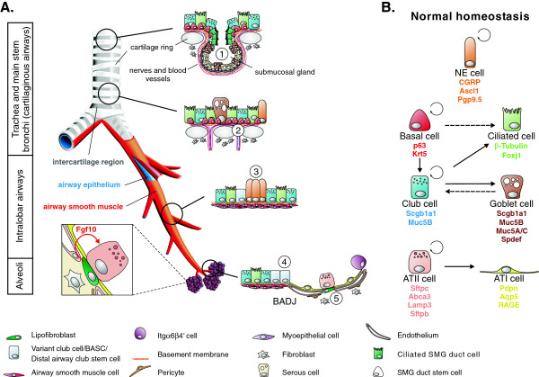
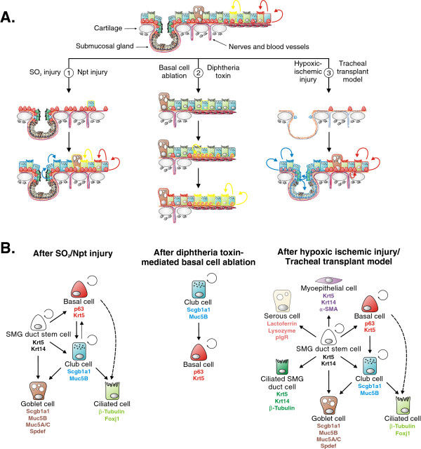
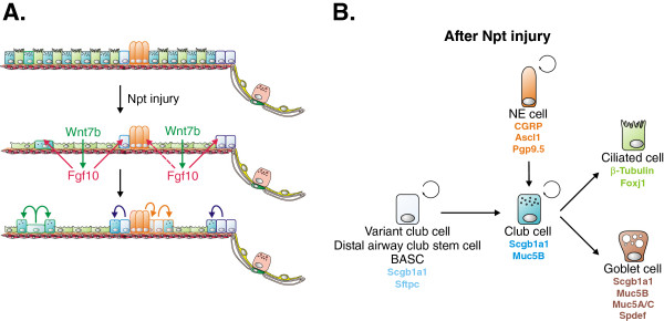
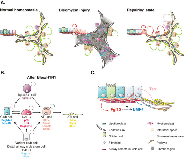
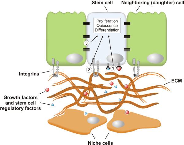
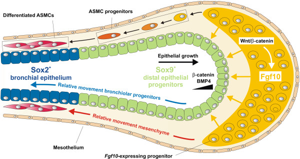
Throughout life adult animals crucially depend on stem cell populations to maintain and repair their tissues to ensure life-long organ function. Stem cells are characterized by their capacity to extensively self-renew and give rise to one or more differentiated cell types. These powerful stem cell properties are key to meet the changing demand for tissue replacement during normal lung homeostasis and regeneration after lung injury. Great strides have been made over the last few years to identify and characterize lung epithelial stem cells as well as their lineage relationships. Unfortunately, knowledge on what regulates the behavior and fate specification of lung epithelial stem cells is still limited, but involves communication with their microenvironment or niche, a local tissue environment that hosts and influences the behaviors or characteristics of stem cells and that comprises other cell types and extracellular matrix. As such, an intimate and dynamic epithelial-mesenchymal cross-talk, which is also essential during lung development, is required for normal homeostasis and to mount an appropriate regenerative response after lung injury. Fibroblast growth factor 10 (Fgf10) signaling in particular seems to be a well-conserved signaling pathway governing epithelial-mesenchymal interactions during lung development as well as between different adult lung epithelial stem cells and their niches. On the other hand, disruption of these reciprocal interactions leads to a dysfunctional epithelial stem cell-niche unit, which may culminate in chronic lung diseases such as chronic obstructive pulmonary disease (COPD), chronic asthma and idiopathic pulmonary fibrosis (IPF).


Figures

Figure 1
Figure 1
The composition of the adult mouse lung epithelium during normal homeostasis. (A) The mouse lung is organized into three anatomical regions. The cartilaginous airways (trachea and main stem bronchi) are lined by a pseudostratified epithelium consisting of secretory (club and goblet), ciliated, basal and a few scattered neuroendocrine (NE) cells. Submucosal glands (SMGs) are located between cartilage rings of the proximal trachea and contain a stem cell population in their ducts (1). Label-retaining basal stem cells are often found in the intercartilage regions (2). The intralobar airway epithelium contains club, ciliated and clusters of NE cells called NE bodies (NEBs), which are often found at branching points. Naphthalene-resistant (variant) club cells are located adjacent to the NEBs (3) and at the bronchioalveolar duct junctions (BADJs) (4), and are presumed to be important for epithelial regeneration. The latter most likely represents a heterogeneous population containing bronchioalveolar stem cells (BASCs) and distal airway club stem cells (DASCs), which are activated after injury. The alveolar epithelium consists mainly of alveolar type (AT) I and ATII cells. The latter is a long-term self-renewing stem cell population also capable of giving rise to ATI cells. Lipofibroblasts in the lung interstitium express Fgf10 and are found juxtaposed to ATII stem cells (5). They are therefore an ideal candidate as a niche that controls the behavior of ATII cells during normal homeostasis and after injury. In addition, the alveoli harbor an alveolar progenitor cell enriched for α6β4 integrins. (B) Lineage relationships of lung epithelial stem cells and their progeny during normal homeostasis. The lung epithelium is maintained by three main stem cell populations: basal cells (cartilaginous airways), club cells (cartilaginous airways and bronchioles) and ATII cells (alveoli). Dashed arrows represent lineage relationships, which are likely to occur but have not yet been definitively established. For details see main text.
Figure 2
Figure 2
Stem cell populations contributing to regeneration of the proximal airway epithelium. (A) Widely used tracheal injury models such as the SO2, the naphthalene (Npt), and the tracheal transplant models, are used to study the contribution of stem cell populations to epithelial regeneration. SO2 and naphthalene injury destroy most luminal cells (1). Although surviving club cells can contribute to epithelial regeneration in the trachea following injury, the majority of newly generated club and ciliated cells arise from activated basal stem cells. A basal cell-like stem cell population residing in submucosal gland (SMG) ducts can also contribute to the regenerative process under these conditions, although they are probably employed to a larger extent after more severe injury. Although basal stem cells are presumed to be at the apex of stem cell hierarchy, club cells have been shown to be able to dedifferentiate and give rise to basal cells after diphtheria toxin-mediated depletion of the basal cell population (2). Club cell-derived basal cells can then give rise to club and ciliated cells during normal homeostasis. A more drastic epithelial injury caused by the loss of blood supply is obtained by the tracheal transplant model, which destroys nearly all epithelial cells except for a few injury resistant basal cells and SMG duct stem cells (3). After blood supply is reestablished, these surviving stem cells can then restore the tracheal surface and SMG epithelium. Colored cell outlines represent lineage trace markers. (B) Lineage relationships of lung epithelial stem cells and their differentiated progeny during regeneration of the tracheal epithelium after SO2/naphthalene injury (left), diphtheria toxin-mediated basal cell depletion (middle) and hypoxic ischemic injury using the tracheal transplant model (right). Dashed arrows represent lineage relationships, which are likely to occur but have not yet been definitively established. For details see main text.
Figure 3
Figure 3
Regeneration of the distal airway epithelium after naphthalene injury. (A) Naphthalene (Npt) injury selectively kills all club cells in the distal conducting airways except for a few naphthalene-resistant club cells located near neuroendocrine (NE) bodies (NEBs) and at the bronchioalveolar duct junction (BADJ). Surviving ciliated cells spread out in an attempt to protect the denuded basement membrane and start to express Wnt7b, which then acts on airway smooth muscle cells to induce proliferation and Fgf10 secretion. Fgf10 acts back on surviving club stem cells to activate them and initiate regeneration. Thus, airway smooth muscle can be regarded as a club stem cell niche. Colored cell outlines represent lineage trace markers. (B) Lineage relationships of lung epithelial stem cells and their differentiated progeny during regeneration of the distal conducting airways after naphthalene injury. For details see main text. BASC, bronchioalveolar stem cell.
Figure 4
Figure 4
Regeneration of alveolar epithelium after injury. (A) Bleomycin-mediated injury results in widespread destruction of all alveolar epithelial cells. Surviving ATII cells are activated following injury, undergo proliferation and restore the alveolar epithelium. The regenerative process after bleomycin injury is associated with the progressive invasion of myofibroblasts, which form fibrotic foci featuring increased extracellular matrix deposition. Several sources for myofibroblasts have been proposed, including epithelial cells, circulating fibroblasts as well as resident (lipo)fibroblasts. Additional distal progenitor cell populations appear to contribute to the regeneration of alveolar epithelium, including Itgα6β4+ cells, Scgb1a1+ cells and distal airway stem cells (DASCs). The latter have basal cell characteristics, most likely originate from distal airway club stem cells and give rise to bronchiolar and alveolar epithelium. Colored cell outlines represent lineage trace markers. (B) Lineage relationships of alveolar epithelial stem cells and their differentiated progeny during regeneration of the alveolar epithelium after bleomycin injury. Dashed arrows represent lineage relationships, which are likely to occur but have not yet been definitively established. (C) Model highlighting molecular crosstalk between distal airway club stem cells and components of their niche, including airway smooth muscle and endothelium. For details see main text. BASC, bronchioalveolar stem cell.
Figure 5
Figure 5
The epithelial stem cell niche. The behavior of epithelial stem cells is regulated by external signals, provided by the microenvironment or niche in which stem cells reside. These signals include growth factors (for example Fgf10) and other stem cell regulatory factors secreted by the niche cells, which can be a wide variety of differentiated cell types, including fibroblasts, smooth muscle cells, endothelial cells, neurons as well as neighboring stem cell progeny (1). Another important component of the stem cell niche is the extracellular matrix (ECM), which acts as a reservoir for growth factors and provides mechanical cues to stem cells, which are translated into biochemical signals through integrins via a process called mechanotransduction (2). Finally, direct cell-cell contact between stem cells and their neighboring progeny, which is mediated by adherens and tight junctions, can also provide essential feedback information to their parent stem cells (3). Integration of these different types of niche signals regulates stem cell activity and behavior such as enhancing stem cell quiescence, promoting transient proliferation or differentiation, and maintaining stem cells in an undifferentiated state.
Figure 6
Figure 6
Fgf10 maintains the distal epithelial progenitor population during early lung development.Fgf10 is expressed in the distal (submesothelial) mesenchyme of the early developing lung and acts on the distal Sox9+ epithelial progenitor cells to maintain them and keep them from acquiring bronchiolar Sox2+ fate. Fgf10 directly activates β-catenin signaling in the distal epithelial progenitor cells, and induces Bmp4 and Sox9 expression. As the epithelial tube grows towards the source of Fgf10 expression, progeny from the distal epithelial progenitor cells leave the distal niche-like environment, assuming a more proximal position along the developing airway and start to express the proximal epithelial marker Sox2. A subset of Fgf10-expressing cells in the distal mesenchyme are progenitors for airway smooth muscle cells (ASMCs) and their amplification, as well as Fgf10 expression, is dependent on mesenchymal Wnt/β-catenin signaling.

Similar articles

Cited by

References

    1. Eisenhauer P, Earle B, Loi R, Sueblinvong V, Goodwin M, Allen GB, Lundblad L, Mazan MR, Hoffman AM, Weiss DJ. Endogenous distal airway progenitor cells, lung mechanics, and disproportionate lobar growth following long-term postpneumonectomy in mice. Stem Cells. 2013;31:1330–1339. doi: 10.1002/stem.1377. - DOI - PMC - PubMed
    1. Rock JR, Randell SH, Hogan BL. Airway basal stem cells: a perspective on their roles in epithelial homeostasis and remodeling. Dis Model Mech. 2010;3:545–556. doi: 10.1242/dmm.006031. - DOI - PMC - PubMed
    1. Rock JR, Onaitis MW, Rawlins EL, Lu Y, Clark CP, Xue Y, Randell SH, Hogan BL. Basal cells as stem cells of the mouse trachea and human airway epithelium. Proc Natl Acad Sci U S A. 2009;106:12771–12775. doi: 10.1073/pnas.0906850106. - DOI - PMC - PubMed
    1. Rock JR, Gao X, Xue Y, Randell SH, Kong YY, Hogan BL. Notch-dependent differentiation of adult airway basal stem cells. Cell Stem Cell. 2011;8:639–648. doi: 10.1016/j.stem.2011.04.003. - DOI - PMC - PubMed
    1. Hong KU, Reynolds SD, Watkins S, Fuchs E, Stripp BR. In vivo differentiation potential of tracheal basal cells: evidence for multipotent and unipotent subpopulations. Am J Physiol Lung Cell Mol Physiol. 2004;286:L643–L649. - PubMed

LinkOut - more resources