

Intrinsic brainstem white epidermoid cyst: An unusual case report
- PMID: 24891906
- PMCID: PMC4040035
- DOI: 10.4103/1817-1745.131487
Intrinsic brainstem white epidermoid cyst: An unusual case report
Abstract
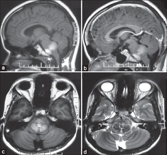
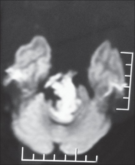
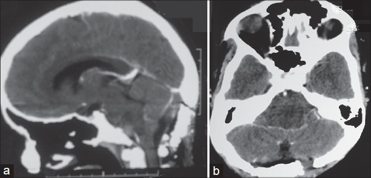
Epidermoid cysts involving the brainstem are extremely rare, with only 18 reported cases in the literature and only five purely intrinsic epidermoid cysts within this group. "White epidermoids", a rare entity, have high protein content and show reversed signal intensity on magnetic resonance images. In contrast to the classical variety, these cysts show high signal intensity on T1-weighted images and low signal intensity on T2-weighted images. Here, we report an interesting case of intrinsic brainstem "white epidermoid cyst" in a 15-year-old girl and discuss its clinical characteristics, radiological features, and surgical treatment. The girl presented with a one-year history of progressive quadriparesis, and features of multiple cranial nerve involvement. Because the cyst was purely intrinsic and had altered signal intensity, the diagnosis was initially unclear until definitive neuroimaging was performed using diffusion-weighted magnetic resonance imaging (DW-MRI) sequences.
Keywords: Brainstem; epidermoid cysts; intrinsic; white.
Conflict of interest statement
Figures
References
-
- Katzman GL. Diagnostic Imaging: Brain. Salt Lake City, Utah: Amirsys; 2004. Epidermoid cyst; p. I-7-16.
-
- Recinos PF, Roonprapunt C, Jallo GI. Intrinsic brainstem epidermoid cyst. Case report and review of the literature. J Neurosurg. 2006;104(Suppl 4):285–9. - PubMed
-
- Berger MS, Wilson CB. Epidermoid cysts of the posterior fossa. J Neurosurg. 1985;62:214–9. - PubMed
-
- Cobbs CS, Pitts LH, Wilson CB. Epidermoid and dermoid cysts of the posterior fossa. Clin Neurosurg. 1997;44:511–28. - PubMed
-
- Toglia JU, Netsky MG, Alexander E., Jr Epithelial (epidermoid) tumors of the cranium. Their common nature and pathogenesis. J Neurosurg. 1965;23:384–93. - PubMed
Publication types
LinkOut - more resources
Full Text Sources
Other Literature Sources
