

High-resolution in vivo imaging of regimes of laser damage to the primate retina
- PMID: 24891943
- PMCID: PMC4033483
- DOI: 10.1155/2014/516854
High-resolution in vivo imaging of regimes of laser damage to the primate retina
Abstract
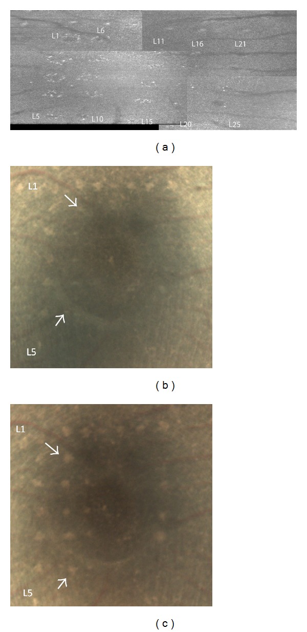
Purpose. To investigate fundamental mechanisms of regimes of laser induced damage to the retina and the morphological changes associated with the damage response. Methods. Varying grades of photothermal, photochemical, and photomechanical retinal laser damage were produced in eyes of eight cynomolgus monkeys. An adaptive optics confocal scanning laser ophthalmoscope and spectral domain optical coherence tomographer were combined to simultaneously collect complementary in vivo images of retinal laser damage during and following exposure. Baseline color fundus photography was performed to complement high-resolution imaging. Monkeys were perfused with 10% buffered formalin and eyes were enucleated for histological analysis. Results. Laser energies for visible retinal damage in this study were consistent with previously reported damage thresholds. Lesions were identified in OCT images that were not visible in direct ophthalmoscopic examination or fundus photos. Unique diagnostic characteristics, specific to each damage regime, were identified and associated with shape and localization of lesions to specific retinal layers. Previously undocumented retinal healing response to blue continuous wave laser exposure was recorded through a novel experimental methodology. Conclusion. This study revealed increased sensitivity of lesion detection and improved specificity to the laser of origin utilizing high-resolution imaging when compared to traditional ophthalmic imaging techniques in the retina.
Figures


Similar articles
-
[Retinal reactions to intense light. I. Threshold lesions. Experimental, morphological and clinical studies of pathological and therapeutic effects of laser and white light].Adv Ophthalmol. 1975;31:159-232. Adv Ophthalmol. 1975. PMID: 810008 German.
-
Subvisible retinal laser therapy: titration algorithm and tissue response.Retina. 2014 Jan;34(1):87-97. doi: 10.1097/IAE.0b013e3182993edc. Retina. 2014. PMID: 23873164
-
High-resolution imaging of the human retina in vivo after scatter photocoagulation treatment using a semiautomated laser system.Ophthalmology. 2010 Mar;117(3):545-51. doi: 10.1016/j.ophtha.2009.07.031. Epub 2010 Jan 19. Ophthalmology. 2010. PMID: 20031226
-
[Pathophysiology of macular diseases--morphology and function].Nippon Ganka Gakkai Zasshi. 2011 Mar;115(3):238-74; discussion 275. Nippon Ganka Gakkai Zasshi. 2011. PMID: 21476310 Review. Japanese.
-
[New examination methods for macular disorders--application of diagnosis and treatment].Nippon Ganka Gakkai Zasshi. 2000 Dec;104(12):899-942. Nippon Ganka Gakkai Zasshi. 2000. PMID: 11193944 Review. Japanese.
Cited by
-
Solar maculopathy: prognosis over one year follow up.BMC Ophthalmol. 2019 Sep 18;19(1):201. doi: 10.1186/s12886-019-1199-6. BMC Ophthalmol. 2019. PMID: 31533669 Free PMC article.
-
Micro-optical coherence tomography of the mammalian cochlea.Sci Rep. 2016 Sep 16;6:33288. doi: 10.1038/srep33288. Sci Rep. 2016. PMID: 27633610 Free PMC article.
-
Non-Therapeutic Laser Retinal Injury.Int J Ophthalmic Res. 2019;5(1):321-335. doi: 10.17554/j.issn.2409-5680.2019.05.90. Epub 2019 Nov 26. Int J Ophthalmic Res. 2019. PMID: 32923732 Free PMC article.
-
Safety assessment in macaques of light exposures for functional two-photon ophthalmoscopy in humans.Biomed Opt Express. 2016 Nov 16;7(12):5148-5169. doi: 10.1364/BOE.7.005148. eCollection 2016 Dec 1. Biomed Opt Express. 2016. PMID: 28018732 Free PMC article.
References
-
- Maiman TH. Stimulated optical radiation in Ruby. Nature. 1960;187(4736):493–494.
-
- Medical Laser Systems-Global Strategic Business Report. Global Industry Analysts. 2013;(365)
-
- Ham WT, Jr., Mueller HA, Ruffolo JJ, Jr., Clarke AM. Sensitivity of the retina to radiation damage as a function of wavelength. Photochemistry and Photobiology. 1979;29(4):735–743. - PubMed
-
- Laser Institute of America. ANSI. American National Standard for Safe Use of Lasers ANSI Z136.1-2014, 2014.
LinkOut - more resources
Full Text Sources
Other Literature Sources
