Review
. 2014 May;3(3):58-69.
doi: 10.7453/gahmj.2014.017.

The Unexpected Uses of Urso- and Tauroursodeoxycholic Acid in the Treatment of Non-liver Diseases

Affiliations
Review

The Unexpected Uses of Urso- and Tauroursodeoxycholic Acid in the Treatment of Non-liver Diseases

Sheila Vang et al. Glob Adv Health Med. 2014 May.

Abstract

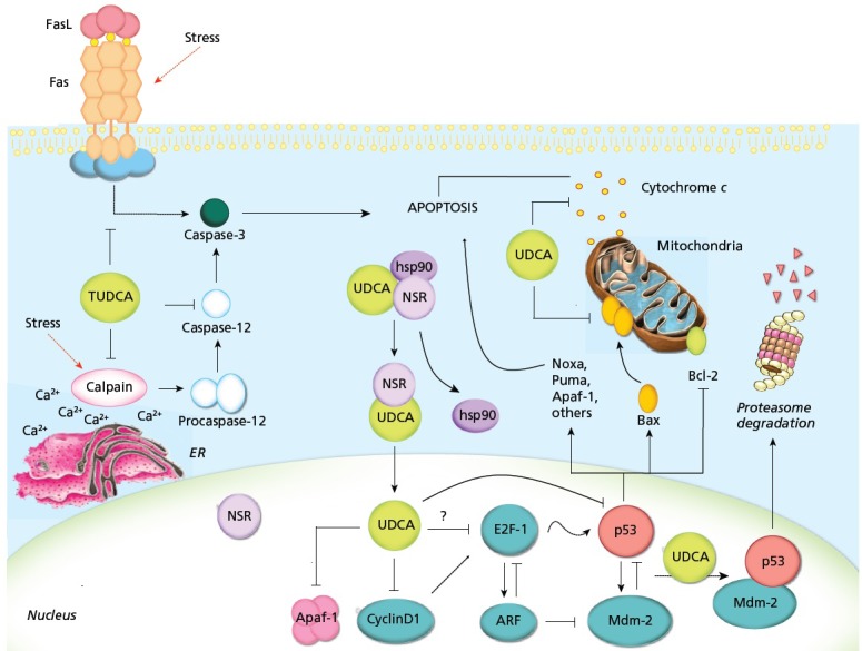
Tauroursodeoxycholic acid (TUDCA) is the taurine conjugate of ursodeoxycholic acid (UDCA), a US Food and Drug Administration-approved hydrophilic bile acid for the treatment of certain cholestatic liver diseases. There is a growing body of research on the mechanism(s) of TUDCA and its potential therapeutic effect on a wide variety of non-liver diseases. Both UDCA and TUDCA are potent inhibitors of apoptosis, in part by interfering with the upstream mitochondrial pathway of cell death, inhibiting oxygen-radical production, reducing endoplasmic reticulum (ER) stress, and stabilizing the unfolded protein response (UPR). Several studies have demonstrated that TUDCA serves as an anti-apoptotic agent for a number of neurodegenerative diseases, including amyotrophic lateral sclerosis, Alzheimer's disease, Parkinson's disease, and Huntington's disease. In addition, TUDCA plays an important role in protecting against cell death in certain retinal disorders, such as retinitis pigmentosa. It has been shown to reduce ER stress associated with elevated glucose levels in diabetes by inhibiting caspase activation, up-regulating the UPR, and inhibiting reactive oxygen species. Obesity, stroke, acute myocardial infarction, spinal cord injury, and a long list of acute and chronic non-liver diseases associated with apoptosis are all potential therapeutic targets for T/UDCA. A growing number of pre-clinical and clinical studies underscore the potential benefit of this simple, naturally occurring bile acid, which has been used in Chinese medicine for more than 3000 years.

Keywords: Apoptosis; bile acids; chaperone; cytoprotection; endoplasmic reticulum stress; misfolded proteins; neurodegenerative diseases; tauroursodeoxy-cholic acid (TUDCA); unfolded protein response; ursodeoxycholic acid (UDCA).


Figures

Figure 1
Figure 1
Proposed mechanisms of T/UDCA inhibition of apoptosis. T/UDCA negatively modulates the mitochondrial pathway by inhibiting Bax translocation, ROS formation, cytochrome c release, and caspase-3 activation. T/UDCA protects against mitochondrial membrane permeabilization and decreased ΔΨm, which reduces ROS production and apoptosis. T/UDCA can also interfere with the death receptor pathway, inhibiting caspase-3 activation. Moreover, T/UDCA inhibits apoptosis associated with ER stress by modulating intracellular calcium levels and inhibiting calpain and caspase-12 activation. Importantly, T/UDCA interacts with NSR, leading to NSR/hsp90 dissociation and nuclear translocation of the T/UDCA/NSR complex. Once in the nucleus, T/UDCA reduces apoptosis by modulating the E2F-1/p53/Bax pathway, inhibiting MDM2/p53 association, decreasing BAX, PUMA and NOXA expression, reducing p53 transactivation and DNA binding activity, and increasing p53 degradation. Finally, T/UDCA downregulates cyclin D1 and Apaf-1, further inhibiting the mitochondrial apoptotic cascade. Abbreviations: Cyt c, cytochrome c; Hsp90, heat shock protein 90; T/UDCA, tauroursodeoxycholic/ursodeoxycholic acid. Reprinted with permission from Amaral et al, 2009.
Figure 2
Figure 2
TUDCA protects striatal mitochondria exposed to 3-NP in vivo. Top left: TUDCA rats exhibit normal striatal mitochondrial size and shape, with intact membrane structure. Bottom left: Enlarged view of TUDCA inset. Top center: Chronic (28 day) 3-NP administration results in abnormal mitochondria. Framed mitochondrion at the same magnification shows dramatic swelling, structural alterations, and loss of membrane integrity. Bottom center: Enlarged view of 3-NP inset. Top right: TUDCA protects mitochondria chronically exposed to 3-NP, as evidenced by normal mitochondrial size, shape, and structure. Bottom right: Enlarged view of 3-NP + TUDCA inset. Magnification is the same for all images. Scale bar, 1 μm. Reprinted with permission from Keene et al, 2001.
Figure 3
Figure 3
Effect of TUDCA on rd10 mouse retinal morphology. Representative micrographs stained with toluidine blue showing significant preservation of photoreceptor nuclei in the outer nuclear layer (ONL) in TUDCA-treated eyes (top panel, right) compared with vehicle-treated eyes (top panel, left). Counts of photoreceptor nuclei were significantly greater in eyes with TUDCA treatment compared to vehicle only. Combined inner segment (IS) and outer segment (OS) length was similarly preserved, with no change in thickness of inner nuclear layer (INL) or ganglion cell layer (GCL). Fluorescence microscopy using a B-2A long pass emission fluorescence filter allows further observation of the preservation of photoreceptor IS and OS present in TUDCA-treated (bottom panel, right) vs vehicle-treated (bottom panel, left) retinal sections. TUNEL-positive nuclei (green/yellow signal) are seen to be abundant in vehicle-treated sections, but are rare in TUDCA-treated sections. TUDCA treatment provided significant preservation of photoreceptor nuclei number in the ONL. Treatment had no discernable effect on the INL or GCL. Reprinted with permission from Boatright et al, 2006.

References

    1. Cash JG, Kuhel DG, Basford JE, et al.Apolipoprotein E4 impairs macrophage efferocytosis and potentiates apoptosis by accelerating endoplasmic reticulum stress. J Biol Chem. 2012;287(33):27876–84 - PMC - PubMed
    1. Amaral JD, Viana RJ, Ramalho RM, Steer CJ, Rodrigues CMP. Bile acids: regulation of apoptosis by ursodeoxycholic acid. J Lipid Res. 2009;50(9):1721–34 - PMC - PubMed
    1. Chakrabarti A, Chen AW, Varner JD. A review of the mammalian unfolded protein response. Biotechnol Bioeng. 2011;108(12):2777–93 - PMC - PubMed
    1. Omura T, Asari M, Yamamoto J, et al.Sodium tauroursodeoxycholate prevents paraquat-induced cell death by suppressing endoplasmic reticulum stress responses in human lung epithelial A549 cells. Biochem Biophys Res Commun. 2013;432(4):689–94 - PubMed
    1. Beuers U. β1 integrin is a long-sought sensor for tauroursodeoxycholic acid. Hepatology. 2013;57(3):867–9 - PubMed