. 2014 Jun 3;9(6):e98623.
doi: 10.1371/journal.pone.0098623. eCollection 2014.

Macrophage colony-stimulating factor augments Tie2-expressing monocyte differentiation, angiogenic function, and recruitment in a mouse model of breast cancer

Affiliations

Macrophage colony-stimulating factor augments Tie2-expressing monocyte differentiation, angiogenic function, and recruitment in a mouse model of breast cancer

Mary A Forget et al. PLoS One. .

Retraction in

Abstract

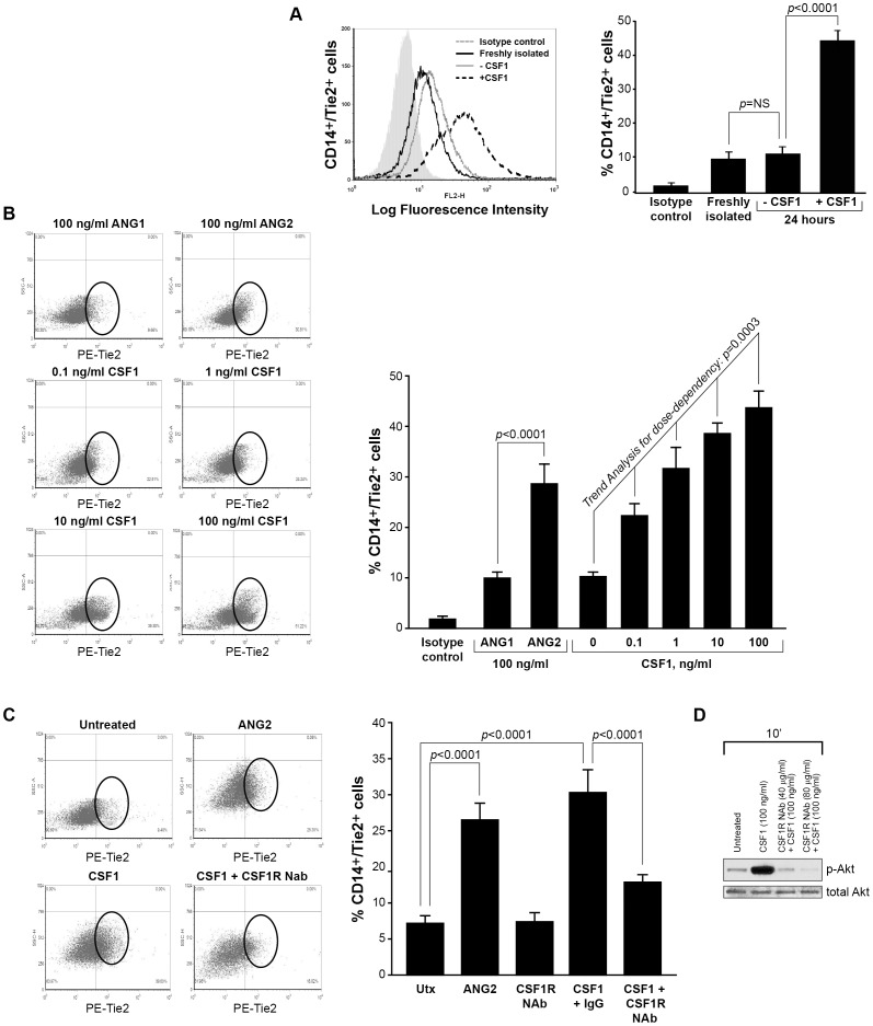
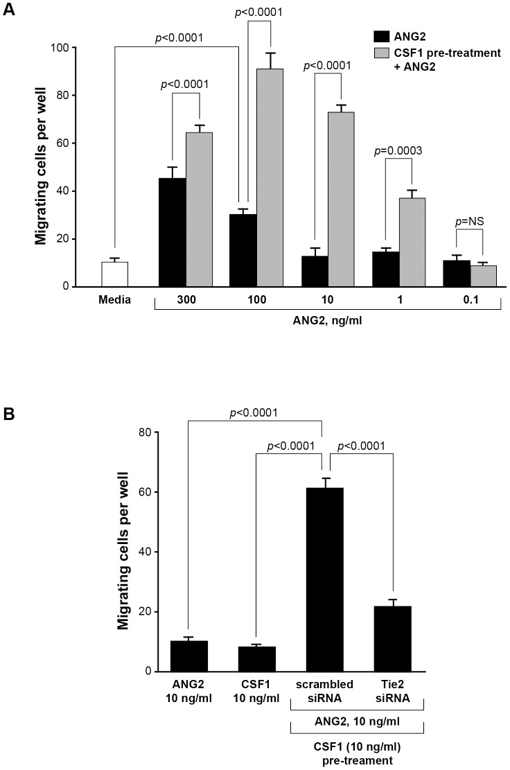
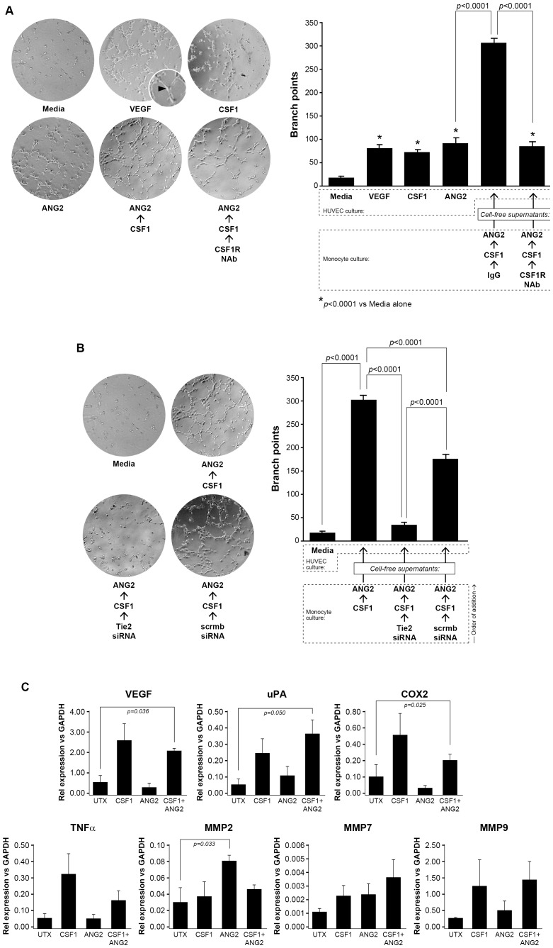
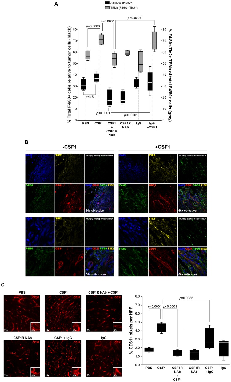
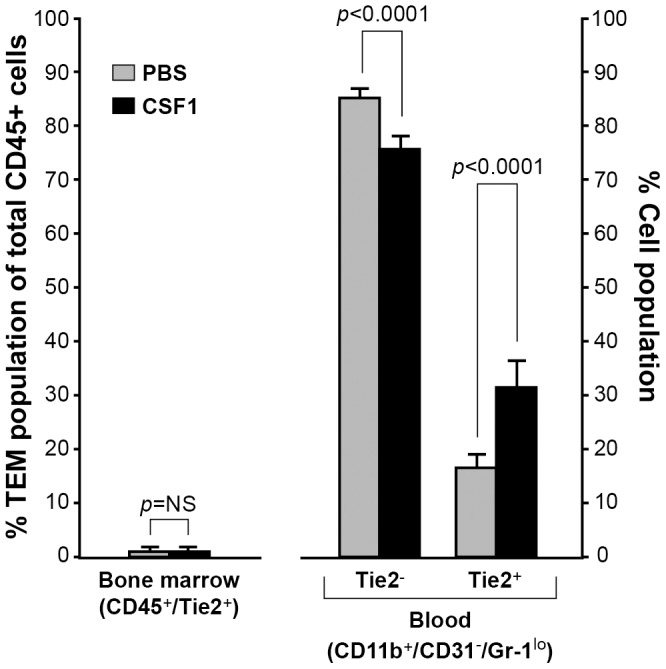
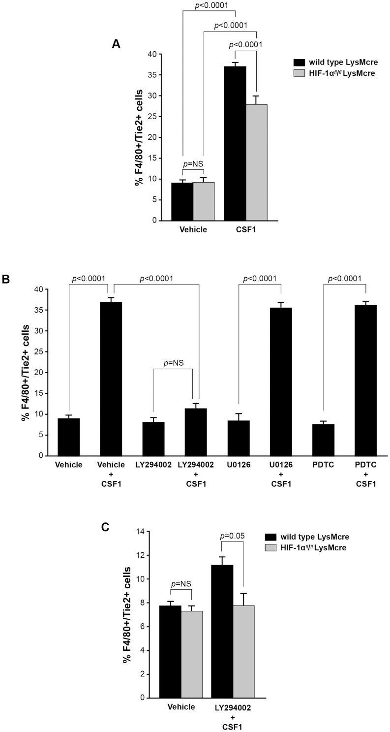
Reports demonstrate the role of M-CSF (CSF1) in tumor progression in mouse models as well as the prognostic value of macrophage numbers in breast cancer patients. Recently, a subset of CD14+ monocytes expressing the Tie2 receptor, once thought to be predominantly expressed on endothelial cells, has been characterized. We hypothesized that increased levels of CSF1 in breast tumors can regulate differentiation of Tie2- monocytes to a Tie2+ phenotype. We treated CD14+ human monocytes with CSF1 and found a significant increase in CD14+/Tie2+ positivity. To understand if CSF1-induced Tie2 expression on these cells improved their migratory ability, we pre-treated CD14+ monocytes with CSF1 and used Boyden chemotaxis chambers to observe enhanced response to angiopoietin-2 (ANG2), the chemotactic ligand for the Tie2 receptor. We found that CSF1 pre-treatment significantly augmented chemotaxis and that Tie2 receptor upregulation was responsible as siRNA targeting Tie2 receptor abrogated this effect. To understand any augmented angiogenic effect produced by treating these cells with CSF1, we cultured human umbilical vein endothelial cells (HUVECs) with conditioned supernatants from CSF1-pre-treated CD14+ monocytes for a tube formation assay. While supernatants from CSF1-pre-treated TEMs increased HUVEC branching, a neutralizing antibody against the CSF1R abrogated this activity, as did siRNA against the Tie2 receptor. To test our hypothesis in vivo, we treated PyMT tumor-bearing mice with CSF1 and observed an expansion in the TEM population relative to total F4/80+ cells, which resulted in increased angiogenesis. Investigation into the mechanism of Tie2 receptor upregulation on CD14+ monocytes by CSF1 revealed a synergistic contribution from the PI3 kinase and HIF pathways as the PI3 kinase inhibitor LY294002, as well as HIF-1α-deficient macrophages differentiated from the bone marrow of HIF-1αfl/fl/LysMcre mice, diminished CSF1-stimulated Tie2 receptor expression.


Conflict of interest statement

Competing Interests: The authors have declared that no competing interests exist.

Figures

Figure 1
Figure 1. CSF1 up-regulates Tie2 receptor on CD14+ human monocytes.
(A) CD14+ monocytes were isolated from whole blood using CD14+ microbeads. Cells were fixed and immunostained using anti-human Tie2 receptor antibody or isotype control antibody immediately following isolation (Freshly isolated) or after treated without (-CSF1) or with rhCSF1 (100 ng/ml) (+CSF1) for 24 hours. N = 10 per group and results represent the mean ± SEM of Tie2-positivity. (B) CD14+ monocytes treated with rhANG1 (100 ng/ml), rhANG2 (100 ng/ml) or a dose-response of rhCSF1 (0, 0.1, 1, 10, 100 ng/ml). ANG2 up-regulated Tie2 expression compared to ANG1 and CSF1 induces a dose-escalation of Tie2 on CD14+ monocytes. N = 10 per group and results represent the mean ± SEM of Tie2-positivity. (C) CD14+ monocytes were left untreated (Utx) or treated with rhANG2 (100 ng/ml) (ANG2), rhCSF1 (100 ng/ml) (CSF1), CSF1R neutralizing antibody alone, or pre-treated with the CSF1R Nab for 30 minutes prior to stimulation with rhCSF1 (100 ng/ml) (CSF1R NAb+CSF1) for 24 hours. ANG2- and CSF1-treatment significantly increased Tie2 expression while the CSF1R NAb abrogated this effect. N = 8 per group and results represent the mean ± SEM of Tie2-positivity by flow cytometry. (D) CD14+ monocytes were left untreated (Untreated), pre-treated with CSF1R NAb (40 µg or 80 µg) for 30 minutes then treated with rhCSF1 (100 ng/ml) (CSF1R NAb+CSF1), or with rhCSF1 (100 ng/ml) alone (CSF1) for 10 minutes. Western blot analysis indicates that the CSF1R NAb was effective at reducing Akt1 phosphorylation.
Figure 2
Figure 2. CSF1 pre-treatment augments the migratory response to ANG2 by CD14+ monocytes.
(A) CD14+ monocytes were isolated and cultured in Boyden chemotaxis chambers in minimal media alone (Media), in media containing 0.1, 1, 10, 100 or 300 ng/ml rhANG2, or with the same ANG2 doses but first pre-treated for 24 hours with media containing 10 ng/ml rhCSF1 and analyzed for their migratory ability. A significant synergistic effect of CSF1 pre-treatment was first observed at 1 ng/ml rhANG2 and peaked at 100 ng/ml rhANG2. N = 8 and results represent the mean ± SEM of CD14+ monocyte migration through the Boyden chamber. (B) CD14+ monocytes were treated with rhANG2 (10 ng/ml) (ANG2) or rhCSF1 (10 ng/ml) (CSF1) alone, or pre-treated with rhCSF1 (10 ng/ml) for 24 hours, washed 3x, then treated with rhANG2 (10 ng/ml) for another 24 hours and transfected with a scrambled siRNA or an siRNA targeting the human Tie2 receptor. While ANG2 and CSF1 did not induce significant migration, the CSF1-pre-treated cells transfected with the scrambled siRNA migrated significantly more than those cells transfected with siRNA targeting Tie2. N = 8 and results represent the mean ± SEM of CD14+ monocyte migration through the Boyden chamber.
Figure 3
Figure 3. Conditioned supernatants from CSF1-treated TEMs augments HUVEC branching.
(A) Human monocytes were isolated from whole blood and differentiated to macrophages over five days using rhCSF1 (5 ng/ml). The cells were serum-starved in endotoxin-free RPMI containing polymyxin B (10 µg/ml) for 24 hours then treated with either a neutralizing antibody for CSF1R or isotype antibody (40 µg/ml) for 1 hour prior to stimulation with CSF1 (10 ng/ml) for 18 hours. The cells were then washed three times and treated with ANG2 (10 ng/ml) for 24 more hours. The conditioned supernatants from these cells (CSF1/ANG2; CSF1R NAb/CSF1/ANG2) or minimal media alone, (minimal), media containing ANG2 alone (10 ng/ml) (ANG2), media containing CSF1 alone (10 ng/ml) (CSF1), or media containing VEGF (10 ng/ml)(VEGF) were used to culture 1.5×10⁴ human umbilical vein endothelial cells (HUVEC) on growth factor-reduced Matrigel for 8 hours. Digital images were captured to determine HUVEC branching. The number of branches was quantified in a blinded manner per field. CSF1, VEGF, and ANG2 each stimulated significantly more branch points from the HUVECs than minimal media. The conditioned media from the macrophages pre-treated with CSF1 induced significantly more branch points than CSF1, VEGF, and ANG2 treatment alone and pre-treatment of the macrophages with the CSF1R neutralizing antibody significantly reduced branch points comparable to media containing VEGF, CSF1, and ANG2 alone levels. N = 5 and results represent the mean ± SEM of HUVEC branch points. (B) In the same manner as above, human monocytes were differentiated to macrophages over five days and serum-starved for 24 hours. The cells were transfected with a siRNA targeting human Tie2 receptor (50 nM) or a scrambled siRNA (50 nM) for 24 hours. The macrophages were washed three times with PBS and then treated with CSF1 (10 ng/ml) for 18 more hours. These conditioned supernatants were collected and used to culture HUVEC cells grown on growth factor-reduced Matrigel for eight hours to detect branch points. Digital images were captured to determine HUVEC branching. The macrophages pre-treated with CSF1 then ANG2 (CSF1/ANG2) induced a significant number of branch points compared to HUVEC cultured in media alone (Media). The conditioned supernatants from the macrophages transfected with siTie2 then treated with CSF1 then ANG2 (Tie2 siRNA/CSF1/ANG2) had significantly less branch points than the CSF1-conditioned supernatants and similar to the minimal conditions. The macrophages transfected with the scrambled siRNA and pre-treated with CSF1 then ANG2 (scrmbsiRNA/CSF1/ANG2) induced significantly more branch points than the conditioned supernatants from the siTie2 samples but still significantly less than the CSF1-conditioned media. N = 5 and results represent the mean ± SEM of HUVEC branch points. (C) CD14+ monocytes were left untreated (UTX) or treated with CSF1 (100 ng/ml) (CSF1), ANG2 (100 ng/ml) (ANG2), or the combination CSF1 (100 ng/ml) and ANG2 (100 ng/ml) (CSF1+ANG2) for 48 hrs. RT-PCR analysis for mRNA expression of the following angiogenic factors was performed: VEGF, uPA, COX2, TNFα, MMP2, MMP7, and MMP9. Data is represented as relative expression for each target mRNA relative to GAPDH housekeeping mRNA. VEGF, uPA, and COX2 mRNA were all significantly increased in CD14+ monocytes treated with CSF1+ANG2 compared to untreated CD14+ monocytes. Each condition was repeated at least 3 times (N = at least 3 and results represent the mean ± SEM for relative mRNA expression).
Figure 4
Figure 4. CSF1 has no effect on tumor growth but increases percent tumor TEMs and augments angiogenesis.
(A) After two weeks of treatment, tumors were removed, homogenized and immunostained with antibodies specific for F4/80 and Tie2 to identify total F4/80+ cells and F4/80+/Tie2+ cells (Tie2-expressing macrophages, TEMs). While there was a marked increase in total F4/80+ macrophages with CSF1 treatment, the percent of F4/80+/Tie2+ TEMs was significantly increased in response to CSF1 suggesting a regulatory role for CSF1 in expanding the TEM population. N = 5 mice per group and results represent the mean ± SEM of total F4/80+ and F4/80+/Tie2+ TEMs within the tumors. (B, top and bottom left) PyMT tumors without CSF1 treatment and (top and bottom right) with CSF1 treatment immunostained with CD31 for blood vessels, F4/80 for macrophages, Tie2 for F4/80+/Tie2+ TEMS, and DAPI. Confocal images (using 60× objective (top) and with 3× zoom (bottom) suggest an increase in both F4/80 macrophages and F4/80+/Tie2+ TEMS in the CSF1-treated tumors. Multiply overlap indicates those areas where F4/80 and Tie2 positivity overlap. Individual stains are in Supplementary Figure 3 . (C, top) Orthotopically implanted PyMT mammary tumors in wild type C57Bl/6 female mice were allowed to become palpable then intraperitoneally treated with PBS (PBS), CSF1 (100 ng in 100 µls) (CSF1), a neutralizing antibody for the CSF1R (50 mg/kg) 4 hours prior to CSF1 treatment (100 ng in 100 µls) (CSF1R NAb+CSF1), the CSF1R antibody alone (CSF1R NAb), an isotype antibody (50 mg/kg) 4 hours prior to CSF1 (100 ng in 100 µls) treatment (CSF1+IgG), or the isotype antibody alone (IgG) three times per week for two additional weeks. The tumors were immunostained with a CD31-Alexa Flour 546 antibody to recognize endothelial cells that comprise blood vessels. Qualitatively, CSF1 treatment increased the percent of CD31-postitive pixels per high powered field compared to PBS treated tumors, while the neutralizing antibody to CSF1R suppressed the CSF1 effect on angiogenesis. (B, bottom) Quantitatively, the percent of CD31+ pixels per high powered field were quantified as blood vessels (angiogenesis) using Adobe Photoshop histogram analysis. CSF1 treatment significantly increased CD31-positive pixels (angiogenesis) compared to PBS. The neutralizing antibody for CSF1R significantly reduced the ability of CSF1 to up-regulate angiogenesis. N = 5 mice per group and results represent the mean ± SEM of percent CD31-positive pixels per high powered field (HPF).
Figure 5
Figure 5. CSF1 expands the TEM population in peripheral blood.
To reach serum levels of CSF1 similar to breast cancer patients and determine that effect on TEM expansion, PBS or CSF1 (20 ng/ml) was intravenously injected into non-tumor bearing wild type C57Bl/6 female mice every other day for a total of three treatments. Bone marrow and atrial blood was collected and immunostained with CD45 and Tie2 antibodies (for bone marrow) or CD11b, CD31, Gr-1, and Tie2 antibodies (for blood). There was no difference in the percentage of TEMs in the bone marrow of the PBS- and CSF1-treated mice. In peripheral blood, CSF1 treatment significantly reduced the percent of CD11b+/CD31+/Gr-1lo/Tie2- cells while significantly increasing CD11b+/CD31+/Gr-1lo/Tie2+ TEMs. N = 5 mice per group and results represent the mean ± SEM of percent total CD45+ and CD45+/Tie2+ cells from bone marrow, and the percent of CD11b+/CD31-/Gr-1lo/Tie2- and CD11b+/CD31-/Gr-1lo/Tie2+ TEMs in peripheral blood.
Figure 6
Figure 6. CSF1 and HIF pathways independently and synergistically regulate Tie2 receptor expression on monocytes.
(A) Bone marrow-derived macrophages were differentiated from age-matched wild type LysMcre and HIF-1αfl/fl/LysMcre mice over five days in non-adherent tubes. After, the cells were serum-starved in endotoxin-free RMPI for 24 hours. The cells were treated with PBS or CSF1 (100 ng/ml) for 24 hours followed by immunostaining with antibodies specific for F4/80 and Tie2 receptor. CSF1 induced an increase in F4/80+/Tie2+ cells over PBS-treated cells from the macrophages derived from the bone marrow of wild type LysMcre mice. The macrophages derived from the HIF-1αfl/fl/LysMcre bone marrow and treated with CSF1 had a significantly smaller percentage of F4/80+ Tie2+ cells than those from the wild type mice. N = 5 per group and results represent the mean ± SEM of percent F4/80+/Tie2+ cells of total F4/80+ cells. (B) Macrophages were derived from the bone marrow of wild type female mice over five days in non-adherent tubes. The cells were serum-starved in endotoxin-free RMPI for 24 hours. The cells were pre-treated with DMSO (vehicle), the PI3 kinase/Akt inhibitor LY294002 (50 µM) (LY294002), the MEK inhibitor U0126 (10 µM) (U0126), or the NF-Kb inhibitor PDTC (100 µM) (PDTC) for 30 minutes. After, the cells were treated with CSF1 (100 ng/ml) (Vehicle+CSF1), LY294002+CSF1 (100 ng/ml) (LY294002+CSF1), U0126+CSF1 (100 ng/ml) (U0126+CSF1), or PDTC+CSF1 (100 ng/ml) (PDTC+CSF1) or left untreated for 24 hours followed by immunstaining with antibodies specific for F4/80 and Tie2. CSF1 induced an increase in the percent of F4/80+/Tie2+ cells compared to vehicle alone. The inhibitors LY294002, U0126, and PDTC alone had no effect on TEM levels. LY294002 pre-treatment significantly reduced the percent of TEMs regulated by CSF1 while U0126 and PDTC had no effect on CSF1 expansion of TEMs. Results represent the mean ± SEM of percent F4/80+/Tie2+ cells of total F4/80+ cells. (C) Bone marrow-derived macrophages were differentiated from age-matched wild type LysMcre and HIF-1αfl/fl/LysMcre mice over five days in non-adherent tubes. After, the cells were serum-starved in endotoxin-free RMPI for 24 hours. The cells were then pre-treated with DMSO or LY294002 (50 µM) for 30 minutes followed by CSF1 (100 ng/ml) (LY294002+CSF1) or not (vehicle) for 24 hours and then immunostained with antibodies specific for F4/80 and Tie2. The macrophages derived from HIF-1αfl/fl/LysMcre mice in combination with the PI3 kinase inhibitor LY294002 significantly reduced the percent TEMs to that similar to untreated levels. N = 5 per group and results represent the mean ± SEM of percent F4/80+/Tie2+ cells of total F4/80+ cells.

Similar articles

Cited by

References

    1. Stanley ER (1986) Action of the colony-stimulating factor, CSF1. Ciba Found Symp. 118: 29–41. - PubMed
    1. Shurtleff SA, Downing JR, Rock CO, Hawkins SA, Roussel MF, et al. (1990) Structural features of the colony-stimulating factor 1 receptor that affect its association with phosphatidylinositol 3-kinase. EMBO J 9: 2415–2421. - PMC - PubMed
    1. Reedijk M, Liu X, van der Geer P, Letwin K, Waterfield MD, et al. (1992) Tyr721 regulates specific binding of the CSF1R kinase insert to PI 3′-kinase SH2 domains: a model for SH2-mediated receptor-target interactions. EMBO J 11: 1365–1372. - PMC - PubMed
    1. Yeung YG, Stanley ER (2003) Proteomic approaches to the analysis of early events in colony-stimulating factor-1 signal transduction. Mol. Cell Proteomics 2: 1143–1155. - PubMed
    1. Curry JM, Eubank TD, Roberts RD, Wang Y, Pore N, et al. (2008) M-CSF signals through the MAPK/ERK pathway via Sp1 to induce VEGF production and induces angiogenesis in vivo. PLoS One 3: e3405. - PMC - PubMed

Publication types

MeSH terms

Substances