. 2014 Jul;28(7):1026-38.
doi: 10.1210/me.2014-1058. Epub 2014 Jun 3.

Naturally occurring mutants inform SHBG structure and function

Affiliations

Naturally occurring mutants inform SHBG structure and function

Tsung-Sheng Wu et al. Mol Endocrinol. 2014 Jul.

Abstract

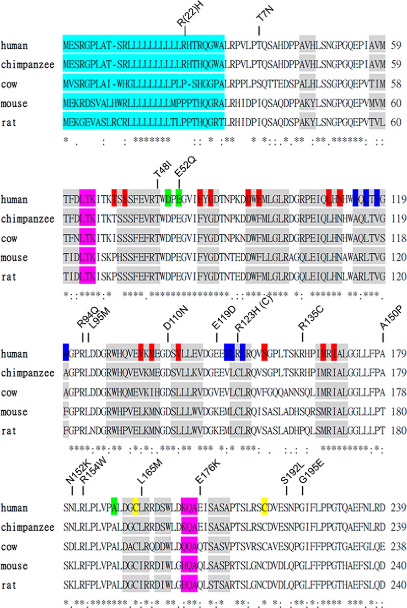
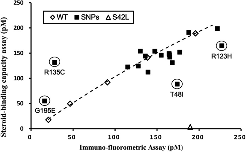
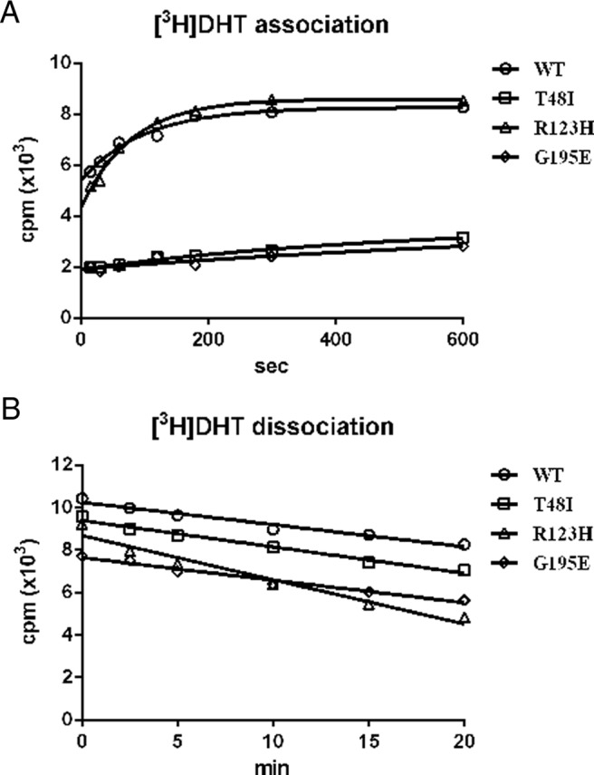
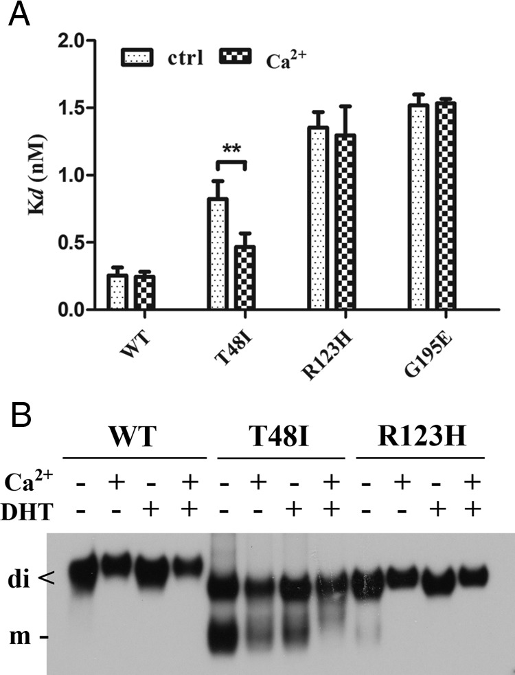
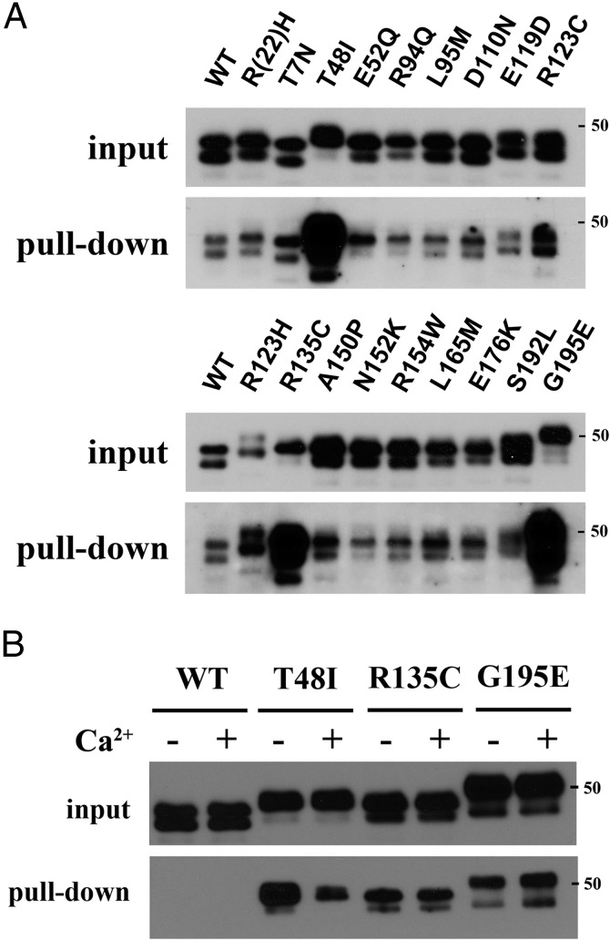
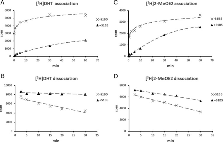
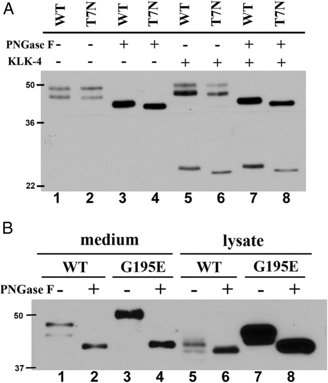
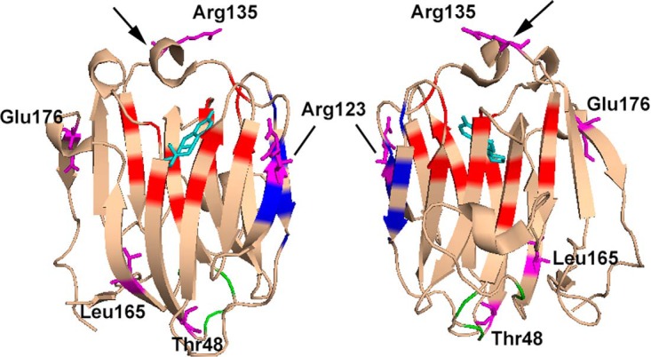
SHBG transports and regulates the activities of androgens and estrogens. Several single nucleotide polymorphisms in the human SHBG gene have been linked to sex steroid-dependent diseases, including those associated with the metabolic syndrome. The N-terminal laminin G-like domain of SHBG includes binding sites for calcium, sex steroids, and fibulin family members, as well as a dimerization domain. We have found that 8 of 18 uncharacterized nonsynonymous single nucleotide polymorphisms within this domain alter the production or biochemical properties of SHBG in ways not previously recognized. O-Linked glycosylation at Thr7 is disrupted in SHBG T7N, whereas abnormal glycosylation of SHBG G195E limits its secretion. Three SHBG mutants (R135C, L165M, and E176K) bind estradiol with abnormally high affinity. SHBG R135C also has an increased interaction with fibulin-2. Two different substitutions within the dimer interface at R123 (R123H and R123C) reduce the affinity for 5α-dihydrotestosterone, while increasing the relative binding affinity for estradiol. SHBG T48I is defective in calcium binding, which leads to a defect in dimerization, reduced affinity for sex steroids, and an enhanced interaction with fibulin-2, which can all be restored by calcium supplementation. These naturally occurring mutants provide insight into SHBG structure and function, and defects in SHBG production or function need to be considered in the context of its utility as a biomarker of diseases.


Figures

Figure 1.
Figure 1.
Orthologous sequence alignment of the SHBG N-terminal LG domain and the amino acid substitutions resulting from selected nonsynonymous SNPs. Amino acid sequences between human and other species of SHBG N-terminal LG domain were aligned. Secondary structures of α-helix and β-strands are shown as pink and gray boxes, respectively. Residues known to be involved in steroid binding (red), dimerization (blue), calcium binding (yellow), and disulfide bridge formation (green) are highlighted. The signal peptide is also highlighted in a light-blue color. Amino acid substitutions are presented on top of the sequences. *, conserved residues between species.
Figure 2.
Figure 2.
Comparison of DHT-binding capacity assay and IFA values of recombinant wild-type and mutant SHBGs. Values for SHBG mutants in which amino acid substitutions introduced by nonsynonymous SNPs (■) are compared with a reference line generated using the values for wild-type SHBG (WT) determined at 5 different concentrations (♢). For reference, the corresponding values for a known DHT binding–deficient mutant, SHBG S42L (5), are shown (▵). Mutants suspected of exhibiting abnormal DHT-binding affinity or immune recognition in the IFA are circled and are positioned below or above the wild-type reference line, respectively.
Figure 3.
Figure 3.
Kinetics of DHT binding to wild-type SHBG (WT) and SHBG mutants with a suspected abnormally low DHT-binding affinity (SHBG T48I, SHBG R123H, and SHBG G195E). A, Association rates were determined by measuring the occupancy of unliganded SHBG samples by [3H]DHT over time at 0°C. B, Dissociation rates were determined in a pulse-chase experiment by preincubating SHBG with 10 nM [3H]DHT for 1 hour followed by adding 3 μM DHT for 0 to 20 minutes at 0°C.
Figure 4.
Figure 4.
Influence of calcium supplementation on SHBG mutants with abnormally low DHT-binding affinity. A, Calcium partially restores the DHT-binding affinity of SHBG T48I but has no effect on other DHT-binding defective SHBG mutants (SHBG R123H and SHBG G195E). Dissociation constants (Kd) of wild-type SHBG (WT), SHBG T48I, SHBG R123H, and SHBG G195E for [3H]DHT were determined by Scatchard analysis in the presence or absence (control) of 1 mM CaCl2. **, P < .01. B, Calcium and/or DHT stabilizes SHBG T48I and SHBG R123H homodimers. Western blotting analysis under nondenaturing conditions was performed in the presence or absence of DHT or calcium using rabbit anti-human SHBG antiserum as the primary antibody.
Figure 5.
Figure 5.
Identification of SHBG mutants with abnormal interactions with fibulin-2. A, Interaction of wild-type SHBG or SHBG mutants with the C-terminal domain of fibulin-2. A GST pull-down assay was performed by using 10 μg of a GST-fibulin-2 fusion protein as bait, and the relative amounts of interacting SHBG were determined by SDS-PAGE and Western blotting using rabbit anti-human SHBG antiserum as the primary antibody. Exposure of the Western blots was adjusted to compare the relative amounts of different SHBG mutants that interacted with fibulin-2 in the GST pull-down assay, against wild-type SHBG as a reference. B, Calcium decreases the interaction of T48I SHBG with fibulin-2 when the GST pull-down assay was performed in the presence or absence of 1 mM CaCl2. Note that this Western blot was underexposed to see the effect of calcium supplementation on fibulin-2 interaction with SHBG mutants with enhanced ability to interact with fibulin-2, and at this exposure the pull down of wild-type SHBG (WT) is not detectable. In all of these Western blots, the denatured recombinant SHBG monomers migrate during SDS-PAGE as a doublet, the sizes and intensities of which reflect differential utilization of the 2 N-linked glycosylation sites in the C-terminal LG domain (14). A 50-kDa molecular size marker is shown on the right of the Western blots, which are representative examples of 3 independent experiments.
Figure 6.
Figure 6.
Effects of S1B5 antibody binding to SHBG on steroid-binding kinetics. Steroid association rates were determined by incubating unliganded wild-type SHBG with or without S1B5 antibody for 1 hour at room temperature followed by incubation (30 seconds to 1 hour at 0°C) with either [3H]DHT (A) or [3H]2-MeOE2 (C). Dissociation rates were determined by first saturating wild-type SHBG with [3H]DHT (B) or [3H]2-MeOE2 (D) followed by an incubation (1 hour at room temperature) in the presence or absence of S1B5 antibody. The irreversible loss of radiolabeled steroids from SHBG over time was then assessed by DCC exposure for 2.5 to 30 minutes at 10°C.
Figure 7.
Figure 7.
Glycosylation status of SHBG T7N and SHBG G195E. A, Lack of glycosylation at Asn7 of SHBG T7N. Wild-type SHBG (WT) or SHBG T7N was treated with PNGase F and/or kallikrein-related peptidase 4 (KLK-4) for 3 hours at 37°C. B, SHBG G195E mostly accumulated in CHO cells with more complexed N-linked glycans. Equal amounts of wild-type SHBG (WT) and G195E SHBG from media or 20 or 5 μg of total cell lysate from CHO cells expressing SHBG wild-type or SHBG G195E were treated with PNGase F at 37°C for 3 hours. Deglycosylated and/or KLK-4 cleaved SHBGs were detected by SDS-PAGE and Western blotting analysis using mouse 7H9 monoclonal antibody. Molecular size markers (kDa) are shown on the left of each Western blot.
Figure 8.
Figure 8.
Crystal structure of the N-terminal LG domain of human SHBG in complex with estradiol (Protein Data Bank code: 1LHU; estradiol is shown in cyan) showing the residues where amino acid substitutions were found to alter the functional properties of SHBG. The positions of residues reported previously (5) to be involved in steroid binding (red), dimerization (blue), and calcium binding (green) are highlighted. The loop structure positioned above the steroid-binding pocket is indicted with an arrow. The residues where substitutions result in altered steroid binding (Thr48, Arg123, Arg135, Leu165, and Glu176), dimerization (Arg123 and Thr48), calcium binding (Thr48), or fibulin-2 binding (Thr48 and Arg135) are shown in purple. Note that Gly195 is not included in this crystal structure of the SHBG N-terminal LG domain.

Comment in

References

    1. Hammond GL. Diverse roles for sex hormone-binding globulin in reproduction. Biol Reprod. 2011;85:431–441. - PMC - PubMed
    1. Hammond GL. Access of reproductive steroids to target tissues. Obstet Gynecol Clin North Am. 2002;29:411–423. - PubMed
    1. Hammond GL, Wu TS, Simard M. Evolving utility of sex hormone-binding globulin measurements in clinical medicine. Curr Opin Endocrinol Diabetes Obes. 2012;19:183–189. - PubMed
    1. Hammond GL, Underhill DA, Smith CL, et al. The cDNA-deduced primary structure of human sex hormone-binding globulin and location of its steroid-binding domain. FEBS Lett. 1987;215:100–104. - PubMed
    1. Grishkovskaya I, Avvakumov GV, Sklenar G, Dales D, Hammond GL, Muller YA. Crystal structure of human sex hormone-binding globulin: steroid transport by a laminin G-like domain. EMBO J. 2000;19:504–512. - PMC - PubMed

Publication types

MeSH terms