Review
. 2014 Jun;124(6):2307-14.
doi: 10.1172/JCI72270. Epub 2014 Jun 2.

Membranous nephropathy: from models to man

Review

Membranous nephropathy: from models to man

Laurence H Beck Jr et al. J Clin Invest. 2014 Jun.

Abstract

As recently as 2002, most cases of primary membranous nephropathy (MN), a relatively common cause of nephrotic syndrome in adults, were considered idiopathic. We now recognize that MN is an organ-specific autoimmune disease in which circulating autoantibodies bind to an intrinsic antigen on glomerular podocytes and form deposits of immune complexes in situ in the glomerular capillary walls. Here we define the clinical and pathological features of MN and describe the experimental models that enabled the discovery of the major target antigen, the M-type phospholipase A2 receptor 1 (PLA2R). We review the pathophysiology of experimental MN and compare and contrast it with the human disease. We discuss the diagnostic value of serological testing for anti-PLA2R and tissue staining for the redistributed antigen, and their utility for differentiating between primary and secondary MN, and between recurrent MN after kidney transplant and de novo MN. We end with consideration of how knowledge of the antigen might direct future therapeutic strategies.


Figures
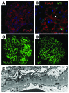
Figure 1
Figure 1. PLA2R staining in normal and MN glomeruli, and EM of typical subepithelial deposits in MN.
(A) IF staining of a normal glomerulus demonstrating PLA2R expression throughout the podocyte (red). Cell nuclei were counterstained with Hoechst dye (blue). (B) A higher-magnification view of a normal glomerulus shows podocytes, labeled with nuclear WT1 (green), that exhibit PLA2R (red) staining diffusely throughout the cell body and processes. The portion of the capillary loop covered by mesangium (arrow) did not stain for PLA2R. In A and B, PLA2R was stained with a polyclonal anti-PLA2R antiserum generated in guinea pig, courtesy of G. Lambeau (Institut de Pharmacologie Moléculaire et Cellulaire, CNRS, and Université de Nice-Sophia Antipolis, Valbonne, France). (C and D) PLA2R staining (green) of a MN kidney biopsy revealed a fine granular capillary loop pattern (C) nearly identical to that of IgG (D). PLA2R was stained with a commercial anti-PLA2R antibody generated in rabbit. (E) EM from a patient with primary MN showed electron-dense deposits (white asterisks) in a subepithelial position beneath the podocyte (P) and overlying the GBM. The podocyte exhibited condensation of the actin cytoskeleton and foot process effacement (arrows) and had laid down new ECM material (black asterisks) between the immune deposits. CL, capillary lumen. Original magnification, ×400 (A, C, and D), ×630 (B).
Figure 2
Figure 2. Mechanisms of subepithelial immune deposit formation.
Left: Circulating antibodies can target surface-exposed intrinsic podocyte proteins to form in situ immune deposits. In PLA2R-associated MN, anti-PLA2R autoantibodies likely bind PLA2R at the podocyte surface to cause capping and shedding of the antigen-antibody complex into the underlying GBM. Middle: Cationized circulating proteins, such as cBSA, may traverse the GBM and bind beneath the podocyte as planted antigens by virtue of their charge, and also serve as the target for circulating antibodies. Right: There is experimental evidence that circulating immune complexes may initially deposit on the luminal side of the GBM, dissociate, and reform in a subepithelial position. Ag, antigen.
Figure 3
Figure 3. Mechanisms of podocyte injury and proteinuria in the passive Heymann nephritis (PHN) model of MN.
(A) The normal podocyte foot process structure is maintained by a well-developed actin cytoskeleton that also serves to anchor the foot processes to the GBM via focal adhesion complexes and cell-matrix adhesion molecules including integrins (α3β1). The normal filtration slit diaphragm (labeled nephrin) forms a final barrier to albumin permeation and is also linked to the actin cytoskeleton via a complex of proteins. (B) In PHN, antibody binding to megalin activates complement leading to assembly and insertion of the MAC (C5b-9). This triggers a cascade of intracellular events (see ref. and the text for details) that contribute to the dissolution of the actin cytoskeleton, which disrupts and displaces the filtration slit diaphragms allowing free passage of albumin into the urine. Collapse of the actin cytoskeleton also affects cell-matrix adhesion and may be the cause of flattening and spreading (effacement) of the podocyte foot processes. Adapted from the American Journal of Physiology — Renal Physiology (90).

References

    1. Polanco N, et al. Spontaneous remission of nephrotic syndrome in idiopathic membranous nephropathy. J Am Soc Nephrol. 2010;21(4):697–704. doi: 10.1681/ASN.2009080861. - DOI - PMC - PubMed
    1. Kidney Disease: Improving Global Outcomes (KDIGO) Glomerulonephritis Work Group KDIGO Clinical Practice Guideline for Glomerulonephritis. Kidney Int Suppl. 2012;2(2):139–274. - PubMed
    1. Border WA, Ward HJ, Kamil ES, Cohen AH. Induction of membranous nephropathy in rabbits by administration of an exogenous cationic antigen. J Clin Invest. 1982;69(2):451–461. doi: 10.1172/JCI110469. - DOI - PMC - PubMed
    1. Schmiedeke TM, Stöckl FW, Weber R, Sugisaki Y, Batsford SR, Vogt A. Histones have high affinity for the glomerular basement membrane. Relevance for immune complex formation in lupus nephritis. J Exp Med. 1989;169(6):1879–1894. doi: 10.1084/jem.169.6.1879. - DOI - PMC - PubMed
    1. Johnson RJ, Couser WG. Hepatitis B infection and renal disease: clinical, immunopathogenetic and therapeutic considerations. Kidney Int. 1990;37(2):663–676. doi: 10.1038/ki.1990.32. - DOI - PubMed

Publication types

MeSH terms