Review
. 2014 Jun;124(6):2355-63.
doi: 10.1172/JCI72269. Epub 2014 Jun 2.

Therapeutic translation in acute kidney injury: the epithelial/endothelial axis

Review

Therapeutic translation in acute kidney injury: the epithelial/endothelial axis

Bruce A Molitoris. J Clin Invest. 2014 Jun.

Abstract

Acute kidney injury (AKI) remains a major clinical event with rising incidence, severity, and cost; it now has a morbidity and mortality exceeding acute myocardial infarction. There is also a documented conversion to and acceleration of chronic kidney disease to end-stage renal disease. The multifactorial nature of AKI etiologies and pathophysiology and the lack of diagnostic techniques have hindered translation of preclinical success. An evolving understanding of epithelial, endothelial, and inflammatory cell interactions and individualization of care will result in the eventual development of effective therapeutic strategies. This review focuses on epithelial and endothelial injury mediators, interactions, and targets for therapy.
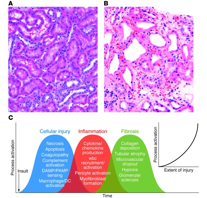
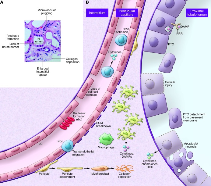
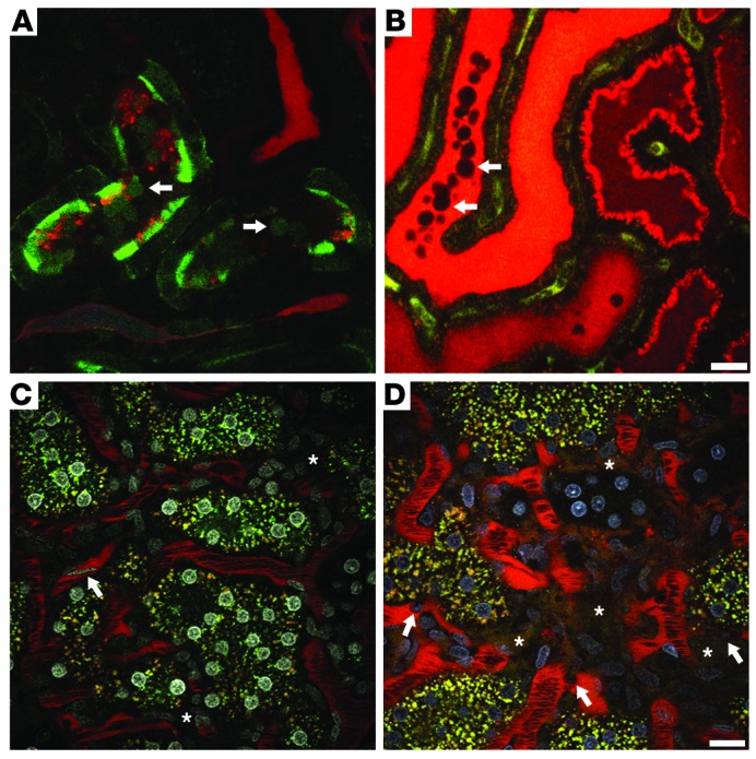

Figures

Figure 1
Figure 1. The complex and overlapping nature of AKI.
(A) Human kidney biopsy with normal cortical morphology. Note the cuboidal PTCs and closely packed tubules surrounded by microvasculature. (B) Human kidney biopsy with ischemic injury (outer stripe). Note the markedly injured PTCs with loss of the apical microvilli and the large number of wbcs occluding flow within the peritubular capillaries, the rouleaux formation, and the marked expansion of the interstitial space. This area of the kidney is especially prone to ongoing vascular congestion after injury, resulting in lack of reperfusion and continuing cellular injury. (C) Multiple cellular injury processes in resident kidney cells lead to local injury and systemic signals for recruitment of circulating wbcs, including PMNs, monocytes, NKTs, Tregs, NK cells, CD4+, CD8+ T cells, and B cells. These cells magnify the ongoing inflammatory process with enhanced resident cell activation (DCs, pericytes, macrophages), cellular dedifferentiation, myofibroblast formation, and ultimately fibrosis and microvascular dropout. The extent of injury determines the level of process activation (inset).
Figure 2
Figure 2. Alterations in the epithelial/endothelial axis during AKI.
(A) Human kidney biopsy with ischemic AKI (cortical area). Note the expanded interstitium, microvascular plugging, dilated tubules, and patchy nature of injury. (B) Under physiologic conditions, coordinated communication and cell-cell interactions maintains homeostasis with normal kidney function. Epithelial injury leads to apoptotic and necrotic cell death that is accompanied by cytokine, chemokine, and ROS release. These factors initiate the release of exogenous and endogenous DAMPS by resident cells, leading to activation and further injury. These signals also initiate the infiltration of professional inflammatory cells such as PMNs and monocytes, leading to enhanced inflammation and further cell injury and destruction. Inflammatory signals produce alterations in the endothelium, resulting in loss of cell-cell contact and breakdown of the ECM. Additionally, there is marked microvascular plugging mediated by adherent wbcs and rouleaux formation, leading to reduced flow and worsening ischemia. Subsequently, there is migration of some of these cells into the interstitium. Pericytes dissociate from underlying ECs and convert into myofibroblasts, which lay down collagen to initiate the fibrotic cycle.
Figure 3
Figure 3. Ischemic injury–induced alterations in the proximal tubules (PTs) and microvasculature.
(A) A portion of the superficial PTs was transfected with GFP-actin using an adenovirus vector. Following i.v. infusion of a small, filterable red dextran, ischemia was induced for 25 minutes, and then blood flow was returned just prior to imaging the area. A single plane from a 3D volume video shows GFP-actin–containing blebs being released from the microvillar membrane (arrows). (B) Lumen of a tubule in cross-section filled with freely filtered red dextran–containing blebs (arrows) flowing down the nephron and forming a cast. The video for B was stitched together from two successively acquired time series. Scale bar: 20 μm. (C) Rhodamine-conjugated dextran (red) is seen circulating within the peritubular microvasculature under physiologic conditions. Flow rates for rbcs, appearing as dark streaks surrounded by bright-red plasma, are high. With the addition of a nuclear dye (white), a fast-flowing wbc can be seen streaking through the vasculature (arrow). Note the absence of red dextran in the interstitial space (asterisks). (D) Rat kidney 24 hours after ischemic injury. Here, activated wbcs with distinguishable Hoechst-labeled nuclei (arrows, nuclei in blue) crawl and roll along the vasculature, slowing down rbc flow. Vessel wall integrity shows heterogeneous areas of damage as the large 150-kDa dextran leaks into the interstitial space (asterisks). The rbc flow rates are markedly reduced due to wbc and rouleaux-mediated obstruction. Scale bar: 20 μm.

Similar articles

Cited by

References

    1. Susantitaphong P, et al. World incidence of AKI: a meta-analysis. Clin J Am Soc Nephrol. 2013;8(9):1482–1493. doi: 10.2215/CJN.00710113. - DOI - PMC - PubMed
    1. Bedford M, Farmer C, Levin A, Ali T, Stevens P. Acute kidney injury and CKD: chicken or egg? Am J Kidney Dis. 2012;59(4):485–491. doi: 10.1053/j.ajkd.2011.09.010. - DOI - PubMed
    1. Coca SG, Singanamala S, Parikh CR. Chronic kidney disease after acute kidney injury: a systematic review and meta-analysis. Kidney Int. 2012;81(5):442–448. doi: 10.1038/ki.2011.379. - DOI - PMC - PubMed
    1. Ishani A, et al. Acute kidney injury increases risk of ESRD among elderly. J Am Soc Nephrol. 2009;20(1):223–228. doi: 10.1681/ASN.2007080837. - DOI - PMC - PubMed
    1. Sharfuddin AA, Molitoris BA. Pathophysiology of ischemic acute kidney injury. Nat Rev Nephrol. 2011;7(4):189–200. doi: 10.1038/nrneph.2011.16. - DOI - PubMed

Publication types

MeSH terms