. 2014 Jun 3:5:4060.
doi: 10.1038/ncomms5060.

Centrin3 in trypanosomes maintains the stability of a flagellar inner-arm dynein for cell motility

Affiliations

Centrin3 in trypanosomes maintains the stability of a flagellar inner-arm dynein for cell motility

Ying Wei et al. Nat Commun. .

Abstract

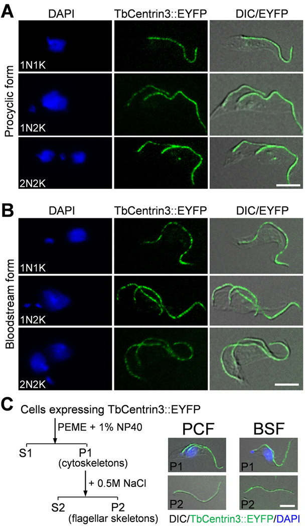
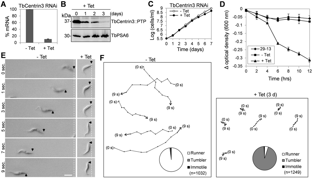
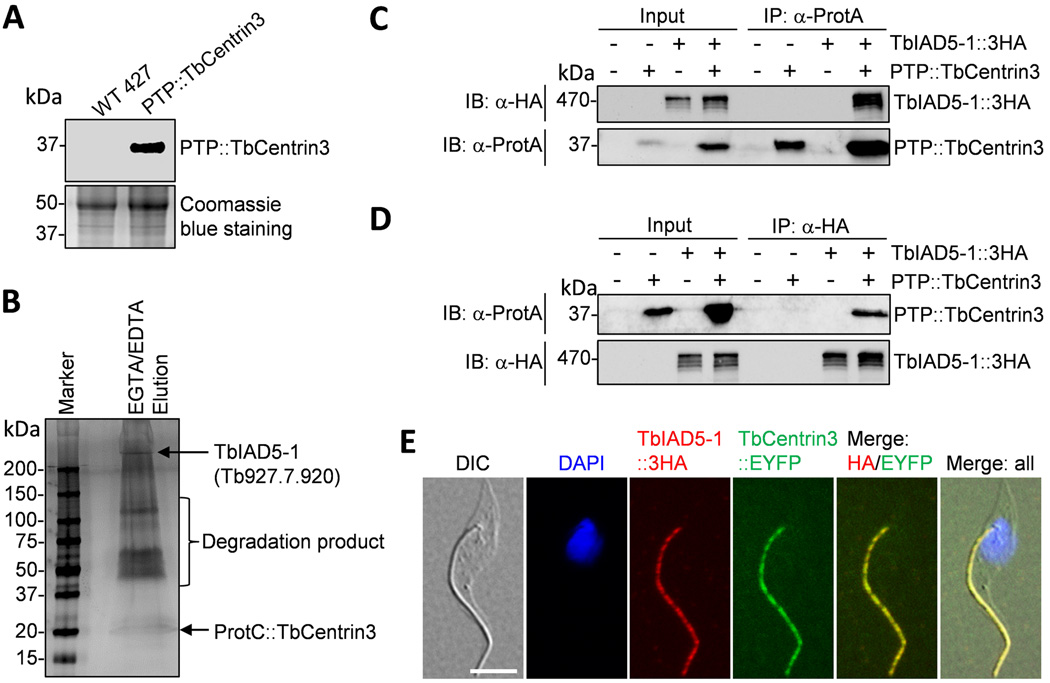
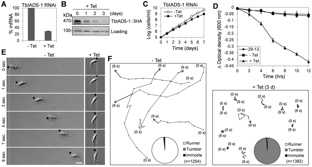
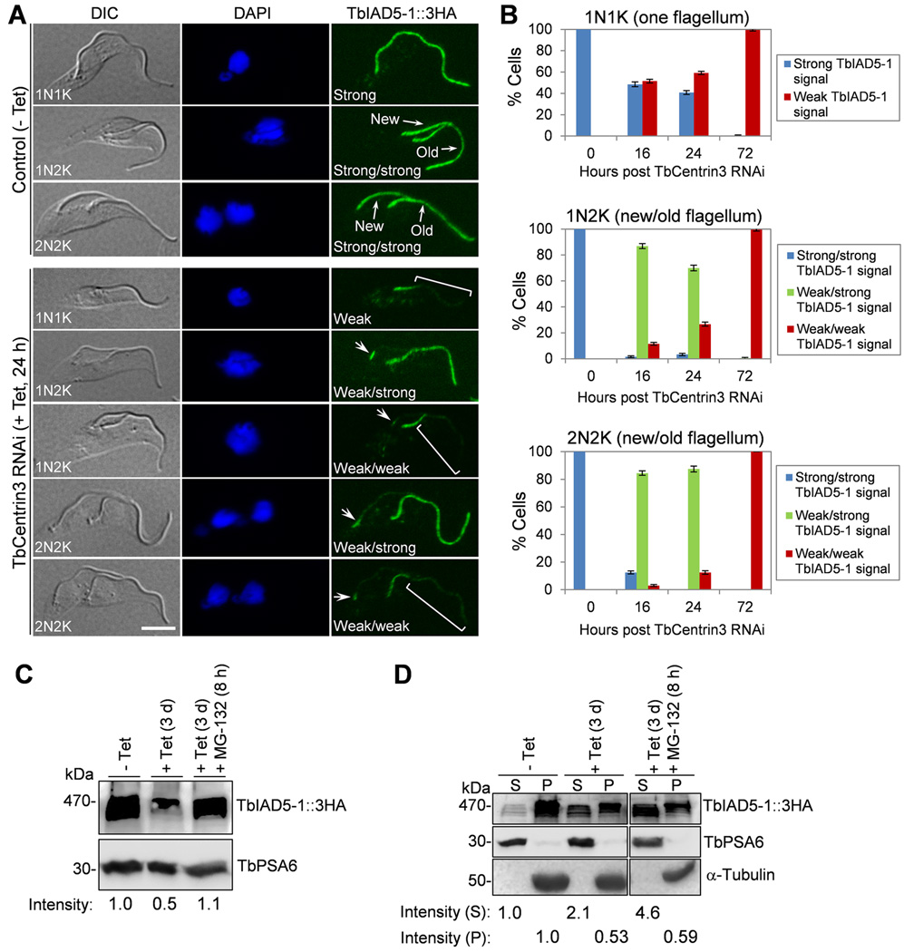
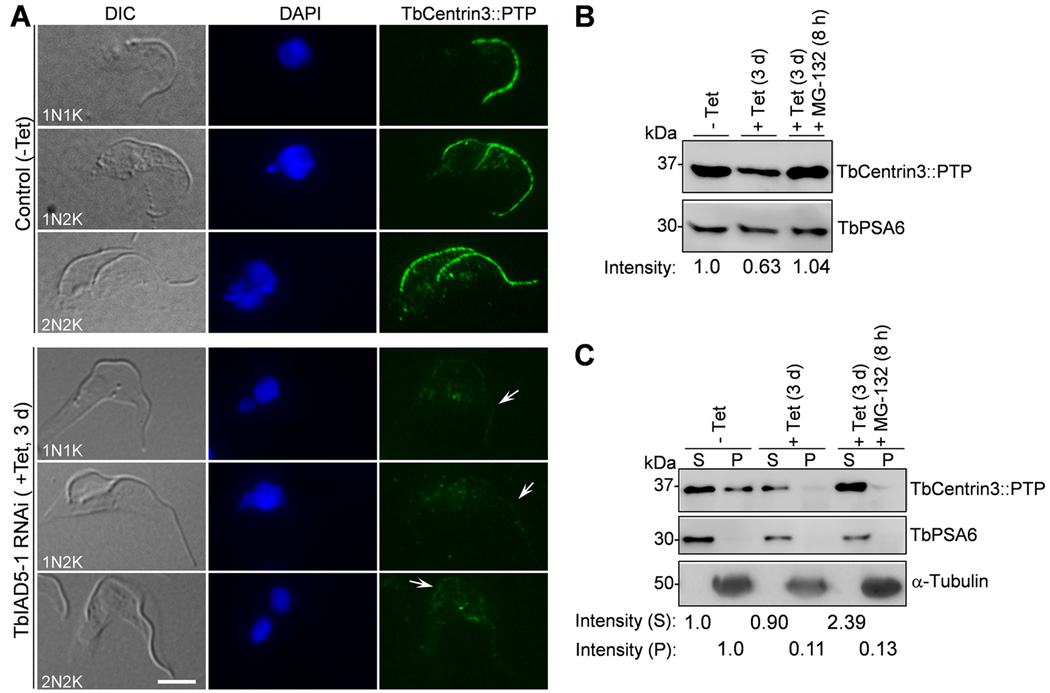
Centrin is a conserved component of centrioles in animals and basal bodies in flagellated organisms. It also associates with axonemal inner-arm dyneins and regulates cell motility, but the underlying mechanism remains elusive. In Trypanosoma brucei, three of the five centrins associate with the flagellar basal body, but no centrin has been found to regulate flagellar motility. Here we show that TbCentrin3 is a flagellar protein and knockdown of TbCentrin3 compromises cell motility. Tandem affinity purification followed by mass spectrometry identifies an inner-arm dynein, TbIAD5-1, as the TbCentrin3 partner, and knockdown of TbIAD5-1 causes similar cell motility defect. Further, we demonstrate the interdependence of TbCentrin3 and TbIAD5-1 for maintaining a stable complex in the flagellar axoneme. Together, these results identify the essential role of TbCentrin3 in cell motility by maintaining the stability of an inner-arm dynein in the flagellum, which may be shared by all the centrin-containing flagellated and ciliated organisms.


Figures

Figure 1
Figure 1. TbCentrin3 is localized to the flagellum in both procyclic and bloodstream forms
(A, B). Localization of TbCentrin3::EYFP in procyclic form (A) and bloodstream form (B). Cells expressing endogenously tagged TbCentrin3::EYFP were treated with PEME buffer containing 1% NP-40, counterstained with DAPI, and visualized under a fluorescence microscope. Bars: 5 µm. (C). TbCentrin3 associates tightly with the flagellum in both the procyclic and bloodstream forms. Cells expressing EYFP-tagged TbCentrin3 were treated with PEME buffer containing 1% NP-40 to prepare cytoskeleton (P1). The cytoskeleton was further treated with PMN buffer containing 1% NP-40 and 500 mM NaCl to prepare flagellar skeleton (P2). The cytoskeleton (P1) and flagellar skeleton (P2) were visualized under the fluorescence microscope. Bar: 5 µm.
Figure 2
Figure 2. RNAi of TbCentrin3 in procyclic trypanosomes impairs cell motility
(A). TbCentrin3 mRNA level in non-induced control cells and RNAi cells detected by quantitative RT-PCR. Three independent experiments were carried out and the error bars represent S.D. (B). TbCentrin3::PTP protein level in control and RNAi cells detected by western blotting with anti-Protein A antibody. Level of TbPSA6, the α6 subunit of the 26S proteasome, was included as the loading control. (C). RNAi of TbCentrin3 slowed down cell proliferation. (D). Sedimentation assays to monitor cell motility. The parental 29-13 strain, the non-induced control, and RNAi cells after tetracycline induction for 3 days were incubated in cuvettes, and the cell density (optical density) was determined and plotted against the time of incubation (hours). Three independent experiments were carried out and the error bars represent S.D. (E, F). Tracing the motility of non-induced control cells and TbCentrin3 RNAi cells (day 3) by video microscopy. Black arrows indicate the posterior tip of the cells, and the white dashed lines show the posterior of cells at the start of time-lapse video microscopy. Bar: 10 µm. Pie charts inserted in panel F show the percentage of cells scored as runner (white), tumbler (gray) or immotile (black) in non-induced control and TbCentrin3 RNAi cells. n: number of cells counted.
Figure 3
Figure 3. TbCentrin3 associates with TbIAD5-1, an inner-arm dynein heavy chain
(A). Western blotting with anti-Protein C antibody to detect the endogenously PTP-tagged TbCentrin3. (B). SDS-PAGE analysis of TbCentrin3-associated proteins. Shown are the final EGTA/EDTA elution of TbCentrin3 PTP purification, which was silver stained. (C). Immunoprecipitation of PTP::TbCentrin3 to precipitate TbIAD5-1::3HA from trypanosome lysate. PTP::TbCentrin3 was detected by anti-Protein A pAb (anti-ProtA), and TbIAD5-1::3HA was detected by anti-HA mAb. (D). Immunoprecipitation of TbIAD5-1::3HA to pull down PTP::TbCentrin3 from trypanosome lysate. (E). Co-localization of TbCentrin3 and TbIAD5-1 in procyclic trypanosomes. Endogenous TbCentrin3 was tagged with EYFP and endogenous TbIAD5-1 was tagged with a triple HA epitope in the same cell line. Cells were immunostained with anti-HA antibody and Cy3-conjugated anti-mouse IgG and counterstained with DAPI. Bar: 5 µm.
Figure 4
Figure 4. RNAi of TbIAD5-1 in the procyclic form causes defects in cell motility
(A). TbIAD5-1 mRNA level in non-induced control and RNAi-induced cells detected by quantitative RT-PCR. Three independent experiments were carried out and the error bars represent S.D. (B). TbIAD5-1::3HA protein level in non-induced control and RNAi cells detected by western blotting with anti-HA antibody. Level of a non-specific protein cross-reacted with anti-HA antibody served as the loading control. (C). RNAi of TbIAD5-1 slowed down cell proliferation. (D). Sedimentation assays to monitor cell motility. The parental 29-13 strain, the non-induced control, and TbIAD5-1 RNAi cells after tetracycline induction for 3 days were incubated in cuvettes, and cell density (optical density) was determined and plotted against the time of incubation (hours). Three independent experiments were carried out and the error bars represent S.D. (E, F). Tracing the motility of non-induced control cells and TbIAD5-1 RNAi cells (day 3) by video microscopy. Black arrows indicate the posterior tip of the cells, and the white dashed lines show the posterior of cells at the start of time-lapse video microscopy. Bar: 10 µm. Pie charts inserted in panel F show the percentage of cells scored as runner (white), tumbler (gray) or immotile (black) in non-induced control and TbIAD5-1 RNAi cells. n: number of cells counted.
Figure 5
Figure 5. Effect of TbCentrin3 knockdown on the localization and stability of TbIAD5-1
(A). TbCentrin3 RNAi on TbIAD5-1 localization to the flagellum. Endogenous TbIAD5-1 was tagged with a C-terminal triple HA epitope in cells harboring the TbCentrin3 stem-loop RNAi construct. TbCentrin3 RNAi was induced by tetracycline for 24 h, and cytoskeleton was prepared for immunostaining with FITC-conjugated anti-HA mAb. New and old flagella are indicated. Arrows in TbCentrin3 RNAi cells indicate the TbIAD5-1 signal at the proximal portion of the flagellum. The square brackets outline the portion of flagellum where TbIAD5-1 signal was reduced. For 1N1K cells, strong or weak signal refers to the TbIAD5-1 signal on the single flagellum. For 1N2K and 2N2K cells, strong/strong signal: strong TbIAD5-1 signal on both new and old flagella; weak/strong signal: weak TbIAD5-1 signal on the new flagellum and strong TbIAD5-1 signal on the old flagellum; weak/weak signal: weak TbIAD5-1 signal on both new and old flagella. Bars: 5 µm. (B) Quantitation of control and TbCentrin3 RNAi cells with different intensity of TbIAD5-1 fluorescence signal. Data are presented as the mean percentage ± S.D. of ~300 cells counted from each of the three independent experiments. All cells from the captured images were counted. (C). Effect of TbCentrin3 RNAi on TbIAD5-1 stability. Crude lysate was analyzed by western blotting with anti-HA mAb to detect 3HA-tagged TbIAD5-1. TbPSA6 level served as the loading control. The intensity of the protein bands was determined with ImageJ, and TbIAD5-1 level was normalized with the loading control. (D). Effect of TbCentrin3 RNAi on TbIAD5-1 protein stability in cytosolic and the cytoskeletal fractions. Non-induced control and TbCentrin3 RNAi cells were lysed in PEME buffer containing 1% NP-40 for cytoskeleton preparation. The soluble cytosolic fraction (S) and cytoskeletal pellet fraction (P) were separated by centrifugation, loaded onto a SDS-PAGE gel, and transferred onto a PVDF membrane for western blotting with anti-HA antibody to detect TbIAD5-1::3HA. The cytosolic fraction was detected by anti-TbPSA6, whereas the cytoskeletal fraction was detected by anti-α-tubulin. The intensity of the protein bands was determined with ImageJ as described above. At least three repeats were carried out.
Figure 6
Figure 6. Effect of TbIAD5-1 knockdown on the localization and stability of TbCentrin3
(A). TbIAD5-1 RNAi on TbCentrin3 localization to the flagellum. Endogenous TbCentrin3 was tagged with a C-terminal PTP epitope in cells harboring the TbIAD5-1 RNAi construct. RNAi was induced by tetracycline for 3 days, and cytoskeleton was prepared for immunostaining with anti-Protein A pAb and FITC-conjugated anti-rabbit IgG. Arrows indicate the weak TbCentrin3::PTP signal at the flagellum upon TbIAD5-1 RNAi. Bar: 5 µm. (B). Effect of TbIAD5-1 RNAi on TbCentrin3 protein stability. Crude lysate was analyzed by western blotting with anti-Protein A pAb to detect PTP-tagged TbCentrin3. Level of TbPSA6 served as the loading control. The intensity of protein bands was determined with ImageJ, and TbCentrin3 level was normalized with the loading control. (C). Effect of TbIAD5-1 RNAi on TbCentrin3 protein stability in cytosolic and the cytoskeletal fractions. Preparation of cytosolic and cytoskeletal fractions was carried out essentially as described in Fig. 5. TbCentrin3::PTP was detected by anti-Protein A pAb. α-Tubulin and TbPSA6 served as the control for cytoskeletal and cytosolic proteins, respectively. The intensity of protein bands was determined with ImageJ and normalized against the loading control. At least three repeats were performed.

Similar articles

Cited by

References

    1. Taillon BE, Adler SA, Suhan JP, Jarvik JW. Mutational analysis of centrin: an EF-hand protein associated with three distinct contractile fibers in the basal body apparatus of Chlamydomonas. J Cell Biol. 1992;119:1613–1624. - PMC - PubMed
    1. Koblenz B, Schoppmeier J, Grunow A, Lechtreck KF. Centrin deficiency in Chlamydomonas causes defects in basal body replication, segregation and maturation. J Cell Sci. 2003;116:2635–2646. - PubMed
    1. Melkonian M. Centrin-mediated cell motility: a novel cell motility mechanism in eukaryotic cells. Bot Acta. 1989;102:3–4.
    1. Geimer S, Melkonian M. Centrin scaffold in Chlamydomonas reinhardtii revealed by immunoelectron microscopy. Eukaryot Cell. 2005;4:1253–1263. - PMC - PubMed
    1. Piperno G, Ramanis Z, Smith EF, Sale WS. Three distinct inner dynein arms in Chlamydomonas flagella: molecular composition and location in the axoneme. J Cell Biol. 1990;110:379–389. - PMC - PubMed

Publication types

MeSH terms

LinkOut - more resources