

Neuroanatomical abnormalities in chronic tinnitus in the human brain
- PMID: 24892904
- PMCID: PMC4148481
- DOI: 10.1016/j.neubiorev.2014.05.013
Neuroanatomical abnormalities in chronic tinnitus in the human brain
Abstract
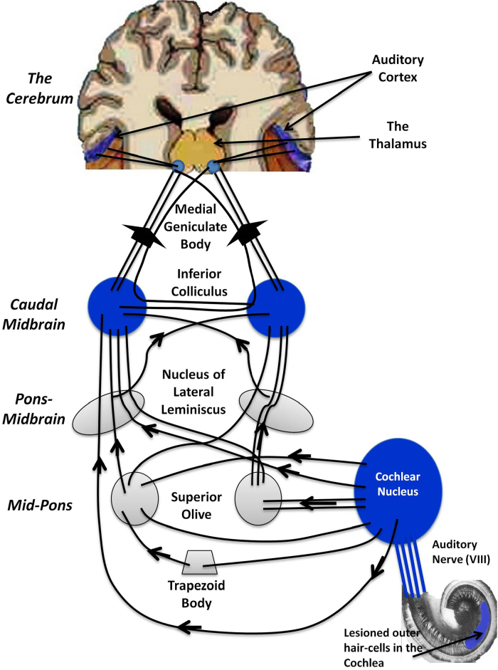
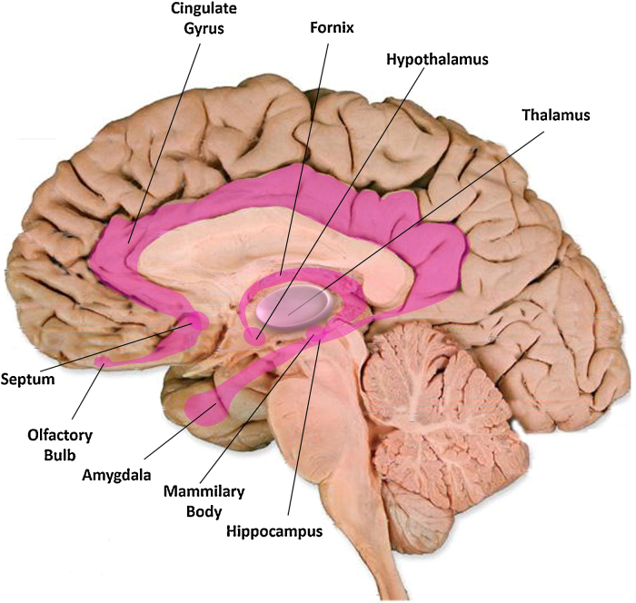
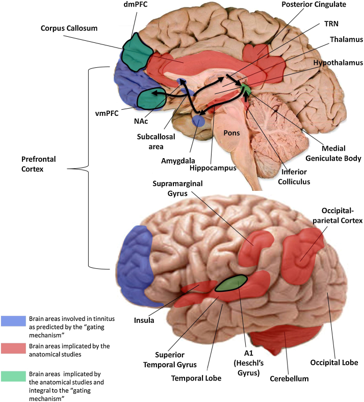
In this paper, we review studies that have investigated brain morphology in chronic tinnitus in order to better understand the underlying pathophysiology of the disorder. Current consensus is that tinnitus is a disorder involving a distributed network of peripheral and central pathways in the nervous system. However, the precise mechanism remains elusive and it is unclear which structures are involved. Given that brain structure and function are highly related, identification of anatomical differences may shed light upon the mechanism of tinnitus generation and maintenance. We discuss anatomical changes in the auditory cortex, the limbic system, and prefrontal cortex, among others. Specifically, we discuss the gating mechanism of tinnitus and evaluate the evidence in support of the model from studies of brain anatomy. Although individual studies claim significant effects related to tinnitus, outcomes are divergent and even contradictory across studies. Moreover, results are often confounded by the presence of hearing loss. We conclude that, at present, the overall evidence for structural abnormalities specifically related to tinnitus is poor. As this area of research is expanding, we identify some key considerations for research design and propose strategies for future research.
Keywords: Gating mechanism; Limbic system; Prefrontal cortex; Tinnitus; Tractography; Voxel-based morphometry.
Copyright © 2014 The Authors. Published by Elsevier Ltd.. All rights reserved.
Figures
References
-
- Adjamian P., Sereda M., Hall D.A. The mechanisms of tinnitus: perspectives from human functional neuroimaging. Hear. Res. 2009;253:15–31. - PubMed
-
- Aldhafeeri F.M., Mackenzie I., Kay T., Alghamdi J., Sluming V. Neuroanatomical correlates of tinnitus revealed by cortical thickness analysis and diffusion tensor imaging. Neuroradiology. 2012;54:889–892. - PubMed
-
- Alexander G.E., Chen K., Merkley T.L., Reiman E.M., Caselli R.J., Aschenbrenner M., Lewis D.J., Pietrini P., Teipel S.J., Hampel H., Rapoport S.I., Moeller J.R. Regional network of magnetic resonance imaging gray matter volume in healthy aging. Neuroreport. 2006;17:951–956. - PubMed
Publication types
MeSH terms
Grants and funding
LinkOut - more resources
Full Text Sources
Other Literature Sources
Medical
