

A proteolytic cascade controls lysosome rupture and necrotic cell death mediated by lysosome-destabilizing adjuvants
- PMID: 24893007
- PMCID: PMC4043491
- DOI: 10.1371/journal.pone.0095032
A proteolytic cascade controls lysosome rupture and necrotic cell death mediated by lysosome-destabilizing adjuvants
Abstract
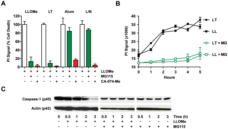
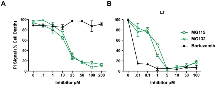
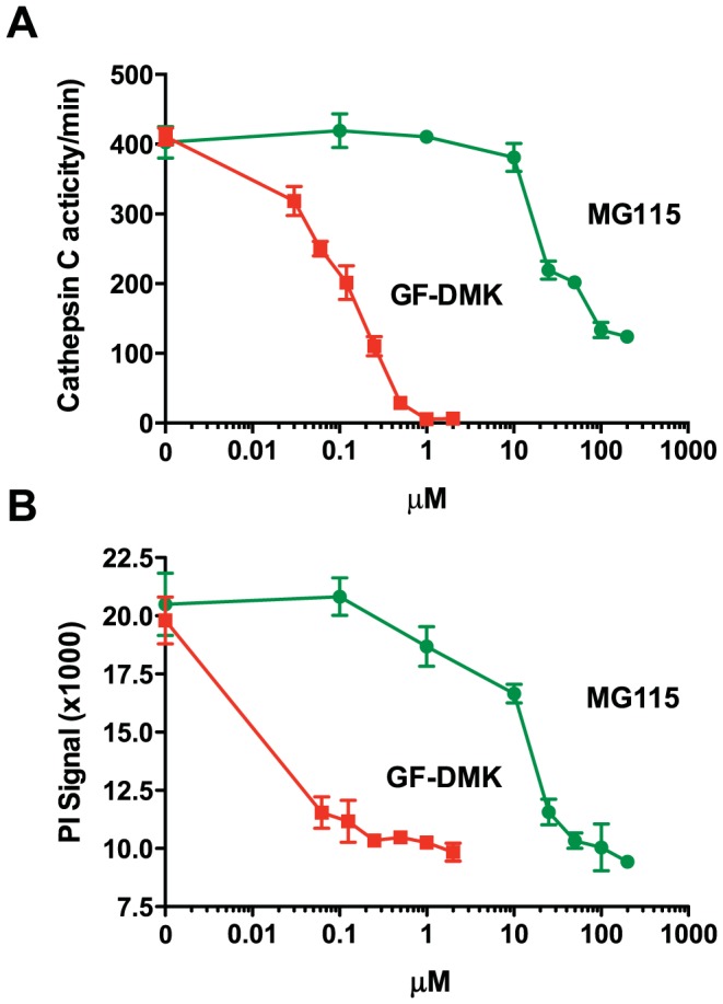
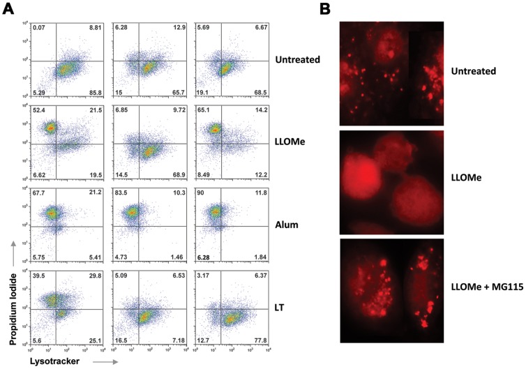
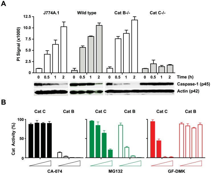
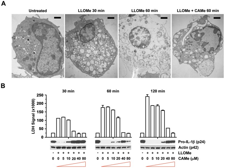
Recent studies have linked necrotic cell death and proteolysis of inflammatory proteins to the adaptive immune response mediated by the lysosome-destabilizing adjuvants, alum and Leu-Leu-OMe (LLOMe). However, the mechanism by which lysosome-destabilizing agents trigger necrosis and proteolysis of inflammatory proteins is poorly understood. The proteasome is a cellular complex that has been shown to regulate both necrotic cell death and proteolysis of inflammatory proteins. We found that the peptide aldehyde proteasome inhibitors, MG115 and MG132, block lysosome rupture, degradation of inflammatory proteins and necrotic cell death mediated by the lysosome-destabilizing peptide LLOMe. However, non-aldehyde proteasome inhibitors failed to prevent LLOMe-induced cell death suggesting that aldehyde proteasome inhibitors triggered a pleotropic effect. We have previously shown that cathepsin C controls lysosome rupture, necrotic cell death and the adaptive immune response mediated by LLOMe. Using recombinant cathepsin C, we found that aldehyde proteasome inhibitors directly block cathepsin C, which presumably prevents LLOMe toxicity. The cathepsin B inhibitor CA-074-Me also blocks lysosome rupture and necrotic cell death mediated by a wide range of necrosis inducers, including LLOMe. Using cathepsin-deficient cells and recombinant cathepsins, we demonstrate that the cathepsins B and C are not required for the CA-074-Me block of necrotic cell death. Taken together, our findings demonstrate that lysosome-destabilizing adjuvants trigger an early proteolytic cascade, involving cathepsin C and a CA-074-Me-dependent protease. Identification of these early events leading to lysosome rupture will be crucial in our understanding of processes controlling necrotic cell death and immune responses mediated by lysosome-destabilizing adjuvants.
Conflict of interest statement
Figures
Similar articles
-
Distinct cathepsins control necrotic cell death mediated by pyroptosis inducers and lysosome-destabilizing agents.Cell Cycle. 2015;14(7):964-72. doi: 10.4161/15384101.2014.991194. Cell Cycle. 2015. PMID: 25830414 Free PMC article.
-
Role of lysosome rupture in controlling Nlrp3 signaling and necrotic cell death.Cell Cycle. 2013 Jun 15;12(12):1868-78. doi: 10.4161/cc.24903. Epub 2013 May 20. Cell Cycle. 2013. PMID: 23708522 Free PMC article.
-
Cathepsin-mediated necrosis controls the adaptive immune response by Th2 (T helper type 2)-associated adjuvants.J Biol Chem. 2013 Mar 15;288(11):7481-7491. doi: 10.1074/jbc.M112.400655. Epub 2013 Jan 7. J Biol Chem. 2013. PMID: 23297415 Free PMC article.
-
Multi‑faceted roles of cathepsins in ischemia reperfusion injury (Review).Mol Med Rep. 2022 Dec;26(6):368. doi: 10.3892/mmr.2022.12885. Epub 2022 Oct 27. Mol Med Rep. 2022. PMID: 36300202 Free PMC article. Review.
-
Engaging the Lysosome and Lysosome-Dependent Cell Death in Cancer.In: Mayrovitz HN, editor. Breast Cancer [Internet]. Brisbane (AU): Exon Publications; 2022 Aug 6. Chapter 13. In: Mayrovitz HN, editor. Breast Cancer [Internet]. Brisbane (AU): Exon Publications; 2022 Aug 6. Chapter 13. PMID: 36122163 Free Books & Documents. Review.
Cited by
-
Bullous Pemphigoid IgG Induces Cell Dysfunction and Enhances the Motility of Epidermal Keratinocytes via Rac1/Proteasome Activation.Front Immunol. 2019 Feb 12;10:200. doi: 10.3389/fimmu.2019.00200. eCollection 2019. Front Immunol. 2019. PMID: 30809225 Free PMC article.
-
Frontline Science: Multiple cathepsins promote inflammasome-independent, particle-induced cell death during NLRP3-dependent IL-1β activation.J Leukoc Biol. 2017 Jul;102(1):7-17. doi: 10.1189/jlb.3HI0316-152R. Epub 2017 Jan 13. J Leukoc Biol. 2017. PMID: 28087651 Free PMC article.
-
Biphasic ROS production, p53 and BIK dictate the mode of cell death in response to DNA damage in colon cancer cells.PLoS One. 2017 Aug 10;12(8):e0182809. doi: 10.1371/journal.pone.0182809. eCollection 2017. PLoS One. 2017. PMID: 28796811 Free PMC article.
-
Phagolysosome acidification is required for silica and engineered nanoparticle-induced lysosome membrane permeabilization and resultant NLRP3 inflammasome activity.Toxicol Appl Pharmacol. 2017 Mar 1;318:58-68. doi: 10.1016/j.taap.2017.01.012. Epub 2017 Jan 24. Toxicol Appl Pharmacol. 2017. PMID: 28126413 Free PMC article.
-
Tumor stressors induce two mechanisms of intracellular P-glycoprotein-mediated resistance that are overcome by lysosomal-targeted thiosemicarbazones.J Biol Chem. 2018 Mar 9;293(10):3562-3587. doi: 10.1074/jbc.M116.772699. Epub 2018 Jan 5. J Biol Chem. 2018. PMID: 29305422 Free PMC article.
References
Publication types
MeSH terms
Substances
Grants and funding
LinkOut - more resources
Full Text Sources
Other Literature Sources
