. 2014 Aug;4(8):956-71.
doi: 10.1158/2159-8290.CD-13-0879. Epub 2014 Jun 3.

EGFR variant heterogeneity in glioblastoma resolved through single-nucleus sequencing

Affiliations

EGFR variant heterogeneity in glioblastoma resolved through single-nucleus sequencing

Joshua M Francis et al. Cancer Discov. 2014 Aug.

Abstract

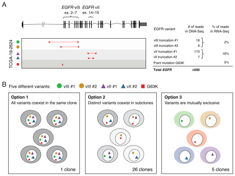
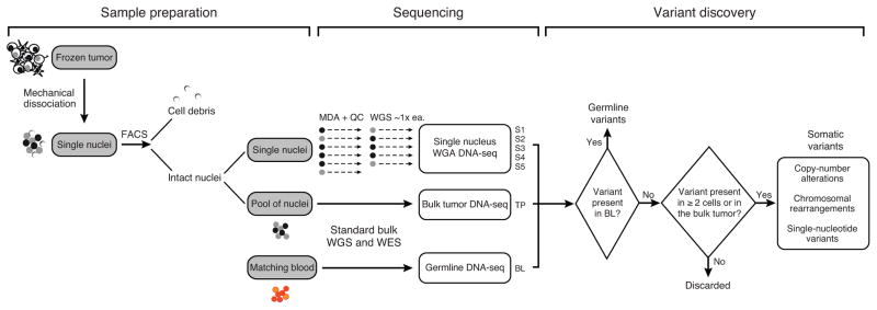
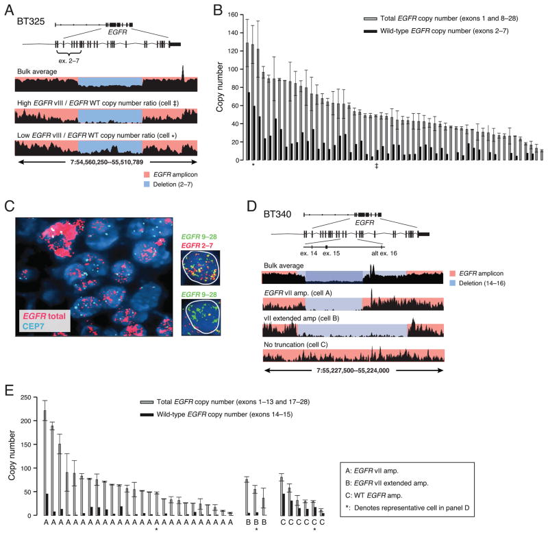
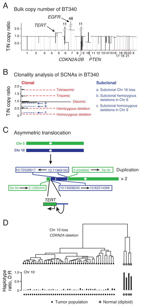
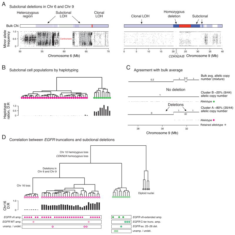
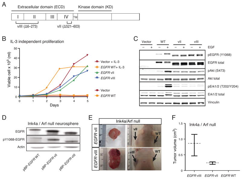
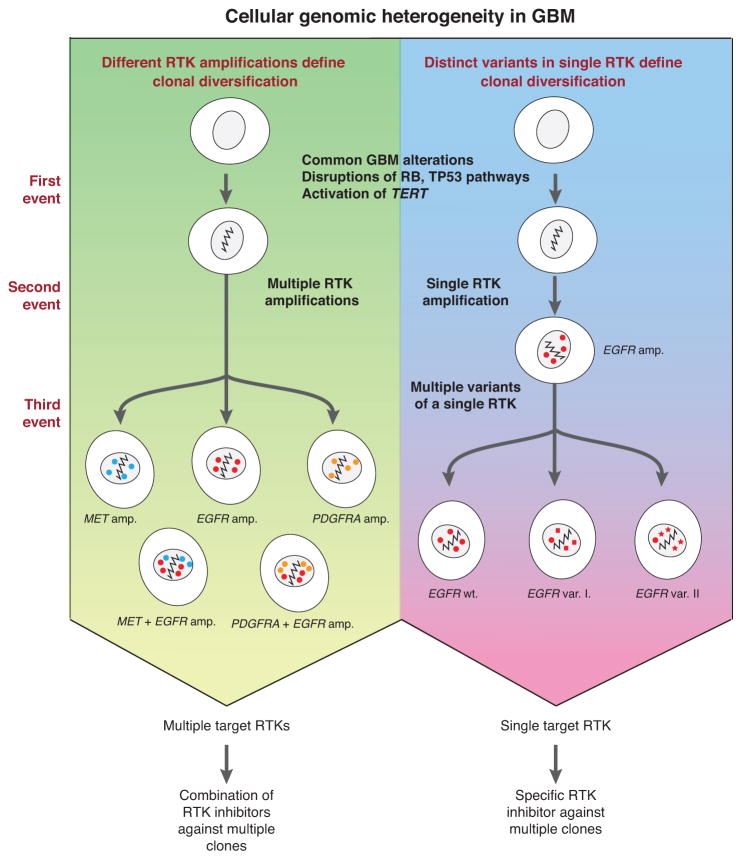
Glioblastomas (GBM) with EGFR amplification represent approximately 50% of newly diagnosed cases, and recent studies have revealed frequent coexistence of multiple EGFR aberrations within the same tumor, which has implications for mutation cooperation and treatment resistance. However, bulk tumor sequencing studies cannot resolve the patterns of how the multiple EGFR aberrations coexist with other mutations within single tumor cells. Here, we applied a population-based single-cell whole-genome sequencing methodology to characterize genomic heterogeneity in EGFR-amplified glioblastomas. Our analysis effectively identified clonal events, including a novel translocation of a super enhancer to the TERT promoter, as well as subclonal LOH and multiple EGFR mutational variants within tumors. Correlating the EGFR mutations onto the cellular hierarchy revealed that EGFR truncation variants (EGFRvII and EGFR carboxyl-terminal deletions) identified in the bulk tumor segregate into nonoverlapping subclonal populations. In vitro and in vivo functional studies show that EGFRvII is oncogenic and sensitive to EGFR inhibitors currently in clinical trials. Thus, the association between diverse activating mutations in EGFR and other subclonal mutations within a single tumor supports an intrinsic mechanism for proliferative and clonal diversification with broad implications in resistance to treatment.

Significance: We developed a novel single-cell sequencing methodology capable of identifying unique, nonoverlapping subclonal alterations from archived frozen clinical specimens. Using GBM as an example, we validated our method to successfully define tumor cell subpopulations containing distinct genetic and treatment resistance profiles and potentially mutually cooperative combinations of alterations in EGFR and other genes.


Conflict of interest statement

Conflict of Interest: M.M. is a founder and equity holder of Foundation Medicine, a for-profit company that provides next-generation sequencing diagnostic services.

Figures

Figure 1
Figure 1. Genomic complexity of the EGFR locus in glioblastoma cannot be resolved using bulk tumor sequencing
(A) RNA-Seq data from GBM patient TCGA-19-2624 revealed co-existence of multiple EGFR aberrations including EGFRvII (16% frequency), EGFRvIII (2% frequency), and a G63K mutation (5% frequency); analysis of whole-genome sequencing further revealed the presence of four distinct intragenic rearrangements producing two variants of EGFRvIII and two variants EGFRvII, each with different allelic frequencies as indicated by read counts. (B) The five distinct EGFR aberrations could result in 25=32 possible clone patterns at the cellular level. Two extreme cases are that all five variants are present in all cells (Option 1) or that the five variants each reside in different cells (Option 3); alternatively, the five variants can exist in up to 26 unique combinations at the cellular level (Option 2).
Figure 2
Figure 2
Experimental and analytical workflow of single-nucleus and bulk tumor sequencing for the characterization of tumor heterogeneity
Figure 3
Figure 3. Heterogeneities in the copy number and mutation status of focally amplified EGFR in two glioblastomas
(A) Read coverage plots of BT325 showing concurrent amplification of EGFR wild type and EGFRvIII alleles (bulk average). Single-nucleus sequencing plots show the same features as bulk but each cell varies in the ratio of EGFR wild-type and EGFRvIII amplification. (B) Quantified levels of total EGFR (exons 1, 8–28) and wild-type EGFR (exons 2–7) across all tumor nuclei using read depths from these regions. EGFRvIII (total minus wild-type) is co-amplified with wild-type in all tumor nuclei but to different degrees. The copy numbers of representative single-nucleus (SN) libraries shown in (A) are denoted by ‡ and ★. (C) Fluorescence in situ hybridization (FISH) for total EGFR and the chromosome 7 centromere (CEP7) confirming varying total EGFR copy number levels in BT325. SureFISH assay (far right nuclei) designed to specifically detect retained and deleted exons shows that individual cells range from low (top) to high levels of vIII deletion (bottom) (D) Read coverage plots of BT340 showing two overlapping deletions encompassing exons 14 and 15 (bulk average). Three representative SN libraries (lower tracks) show different mutation patterns. Cell A contained only EGFRvII truncation, cell B contained only the longer EGFRvII (vII-extended), and Cell C contained only wild-type EGFR. (E) Quantified levels of total EGFR (exons 1–13, 17–28) and wild-type EGFR (exons14–15) read depth within single tumor nuclei demonstrate three distinct populations containing amplified EGFRvII, EGFRvII-extended or wild-type EGFR. Discordant read pair signatures confirmed the presence of vII or vII-extended variants in population A and B; no discordant pairs were found in population C. Asterisks denote the representative nuclei displayed in (D).
Figure 4
Figure 4. Characterization of clonal genomic alterations in single tumor nuclei from BT340
(A) Bulk average somatic copy-number profile derived from whole-genome sequencing of GBM BT340 showing characteristic glioblastoma alterations. T/N= tumor vs. normal. (B) Clonality of somatic copy-number alterations inferred from the copy ratios (18). Clonal SCNAs generate integral copy-number alterations with peaks of copy ratios separated by equal spacing (red dashed lines); off-peak copy ratios corresponding to non-integral copy-number alterations suggest subclonal SCNA events (blue dashed lines). The clonality of subclonal SCNA events was later resolved at the single-cell level (see Fig. 5). (C) Schematic drawing of the inter-chromosomal rearrangements between Chr. 5 and Chr. 10 leading to TERT rearrangement and duplication. Translocation fuses a super enhancer element in Chr. 10 to the 5′ promoter region of TERT. (D) Hierarchical clustering of 48 nuclei from BT340 based on the haplotypes in chromosome 10. Forty-four nuclei (black circles) harbored loss-of-heterozygosity in Chr. 10 (with almost none of the deleted haplotype) and four nuclei (open circles) as heterozygous in Chr. 10 (1:1 ratio between the deleted and the retained haplotypes). The same two groups were also validated by clustering using other clonal aberrations (Supplementary Fig. 6).
Figure 5
Figure 5. Subclonal deletions delineate two tumor populations harboring distinct EGFR variants
(A) Subclonal LOH in chromosomes 6 and 9 of BT340 is reflected by incomplete segregation of SNVs from the population of single tumor nuclei. Gray masking on the left panel indicates regions of subclonal LOH flanked by regions with clonal LOH in chromosome 6. Regions of subclonal homozygous deletions in chromosome 9 have a few heterozygous sites but there are none in regions of clonal homozygous deletion. (B) Hierarchical clustering of 44 tumor nuclei from BT340 based on the single-nucleus haplotypes in regions of subclonal LOH in Chr. 6 identifies two clusters with either loss or preservation of heterozygosity. (C) Two segments in 9p21 with bulk average copy number ~0.20 (top track) are resolved as a mixture of one subclone with 80% of tumor nuclei (35/44) having deletions in both regions. Gray dots show the retained haplotype that is only present in regions that are not deleted. (D) Correlation between subclonal populations as delineated by deletions in 6p11.2, 6q12 and 9p21.1 and distinct EGFR truncations suggest that the two distinct EGFRvII truncations independently emerged after clonal segregation determined by the deletion events.
Figure 6
Figure 6. EGFRvII transforms Ba/F3 and Ink4a/Arf−/− neural stem cells
(A) Schematic representation of the EGFR vII and vIII mutants. (B) Proliferation of Ba/F3 cells stably containing empty vector or expressing EGFR wild-type, EGFRvII, or EGFRvIII were plated in the presence or absence of IL-3. Proliferation was measured over 5 days. (C) Immunoblot of Ba/F3 cells stably expressing empty vector, EGFR wild-type, EGFRvII or EGFRvIII. Cells were serum starved for 3 hours and then stimulated with 25 ng/ml of EGF for 10 min. (D) Immunoblot of Ink4a/Arf null neurospheres stably expressing EGFR wild-type, EGFRvII or EGFRvIII. (E) Xenografted Ink4a/Arf null neurospheres stably expressing EGFRvII or EGFRvIII form subcutaneous tumors after 6- or 11-weeks, respectively. (F) Measurement of the tumor volumes of subcutaneous xenografts stably expressing EGFRvII or EGFRvIII after 6- or 11-weeks respectively. Cells expressing wild-type EGFR failed to form tumors.
Figure 7
Figure 7. Summary of cellular genomic heterogeneity in glioblastoma
(Left panel) Different receptor-tyrosine kinase amplifications can lead to clonal diversification after common clonal mutations. (Right panel) Mutations in a single receptor-tyrosine kinase can also lead to clonal diversification through multiple variants, SNVs, and amplification of these different alleles. The presence of multiple concurrent RTK amplifications/mutations implies potential need for multiple drugs targeting the same RTK via different mechanisms.

Comment in

References

    1. Furnari FB, Fenton T, Bachoo RM, Mukasa A, Stommel JM, Stegh A, et al. Malignant astrocytic glioma: genetics, biology, and paths to treatment. Genes Dev. 2007;21:2683–710. - PubMed
    1. TCGA. Comprehensive genomic characterization defines human glioblastoma genes and core pathways. Nature. 2008;455:1061–8. - PMC - PubMed
    1. Parsons DW, Jones S, Zhang X, Lin JC, Leary RJ, Angenendt P, et al. An integrated genomic analysis of human glioblastoma multiforme. Science. 2008;321:1807–12. - PMC - PubMed
    1. Brennan CW, Verhaak RG, McKenna A, Campos B, Noushmehr H, Salama SR, et al. The somatic genomic landscape of glioblastoma. Cell. 2013;155:462–77. - PMC - PubMed
    1. Nathanson DA, Gini B, Mottahedeh J, Visnyei K, Koga T, Gomez G, et al. Targeted therapy resistance mediated by dynamic regulation of extrachromosomal mutant EGFR DNA. Science. 2014;343:72–6. - PMC - PubMed

Publication types