Review
. 2014 Jun 3:9:106.
doi: 10.1186/1746-1596-9-106.

Giant pelvic angiomyofibroblastoma: case report and literature review

Affiliations
Review

Giant pelvic angiomyofibroblastoma: case report and literature review

Ping Qiu et al. Diagn Pathol. .

Abstract

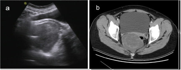
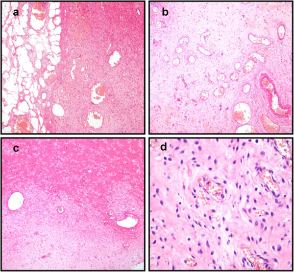
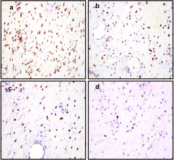
Angiomyofibroblastoma (AMF) is a rare, benign, soft-tissue tumor, which predominantly occurs in the vulvovaginal region of middle-aged women. It is clinically important to distinguish an AMF from other stromal cell lesions. Here, we report the case of a 32-year-old woman with a rare, giant pelvic AMF, which showed a benign clinical course. The tumor was located in the cul-de-sac of Douglas. It was well demarcated, hypocellular, edematous and composed of spindle-shaped and oval stromal cells aggregating around thin-walled blood vessels. The tumor cells had abundant eosinophilic cytoplasm, and expressed estrogen receptors, progesterone receptors and desmin. Mitotic figures were absent. It is important to distinguish AMFs from aggressive angiomyxomas because both occur at similar sites but show different clinical behaviors. Most AMFs and aggressive angiomyxomas have the same immunohistochemical phenotype. The well-circumscribed borders of AMF are the most important characteristic that distinguish it from aggressive angiomyxomas. AMFs rarely recur after complete surgical excision.

Virtual slides: The virtual slide(s) for this article can be found here: http://www.diagnosticpathology.diagnomx.eu/vs/5510813471244189.


Figures

Figure 1
Figure 1
Image feature of tumor pre-operation. (a) Transvaginal ultrasonography shows an oval, moderately echoic mass in close proximity to the cul-de-sac of Douglas. (b) Pelvic CT scan shows a well-circumscribed tumor of homogeneous intensity located anteroinferior to the sacrum.
Figure 2
Figure 2
Histopathological examination of tumor post-operation. (a) The tumor is well demarcated from the surrounding fat tissues. (b) Abundant thin-walled blood vessels can be seen in the tumor. (c) The stroma of the tumor is hyalinized or edematous, and appears hypocellular in some areas. (d) The tumor is composed of bland, plump, spindle-shaped or oval cells that are frequently aggregated around thin-walled blood vessels (H&E: 100 x).
Figure 3
Figure 3
Immunostaining of the tumor cells. (a) The tumor cells were positive for desmin. (b) The tumor cells were immunoreactive with estrogen receptor (c) and progesterone receptor. (d) The tumor cells showed a very low Ki-67 index (IHC: 100 x).

References

    1. Fletcher CD, Tsang WY, Fisher C, Lee KC, Chan JK. Angiomyofibroblastoma of the vulva. A benign neoplasm distinct from aggressive angiomyxoma. Am J Surg Pathol. 1992;16:373–382. doi: 10.1097/00000478-199204000-00006. - DOI - PubMed
    1. Babala P, Biro C, Klacko M, Miklos P, Ondrus D. Angiomyofibroblastoma of the cervix uteri: a case report. Klin Onkol. 2011;24:133–136. - PubMed
    1. Mortele KJ, Lauwers GJ, Mergo PJ, Ros PR. Perineal angiomyofibroblastoma: CT and MR findings with pathologic correlation. J Comput Assist Tomogr. 1999;23:687–689. doi: 10.1097/00004728-199909000-00007. - DOI - PubMed
    1. Naheed S, Upadhyay K, Pradeep K. Angiomyofibroblastoma of the vulva. J Obstet Gynaecol. 2011;31:554–555. doi: 10.3109/01443615.2011.581314. - DOI - PubMed
    1. Lee SH, Yang JW, Do JM, Seo DH, Jung JH, Chung KH, Lee JS, Hyun JS. Angiomyofibroblastoma-like tumor of the scrotum. Korean J Urol. 2010;51:365–367. doi: 10.4111/kju.2010.51.5.365. - DOI - PMC - PubMed

MeSH terms

Substances

LinkOut - more resources