Review
. 2014 Jul;15(7):431-42.
doi: 10.1038/nrn3723. Epub 2014 Jun 4.

Müller glial cell reprogramming and retina regeneration

Affiliations
Review

Müller glial cell reprogramming and retina regeneration

Daniel Goldman. Nat Rev Neurosci. 2014 Jul.

Abstract

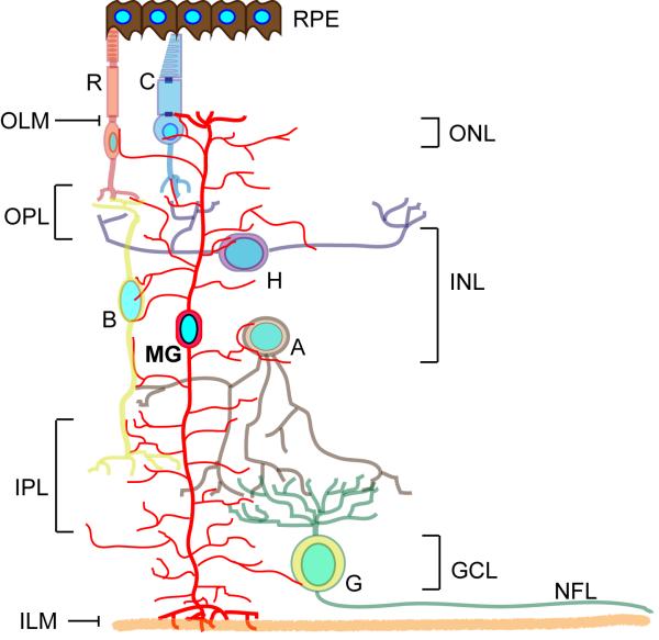
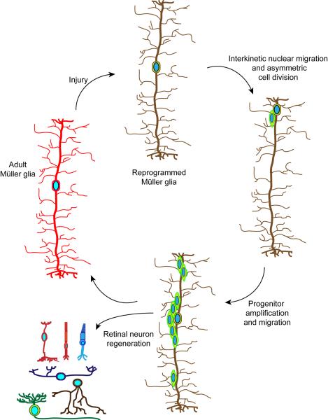
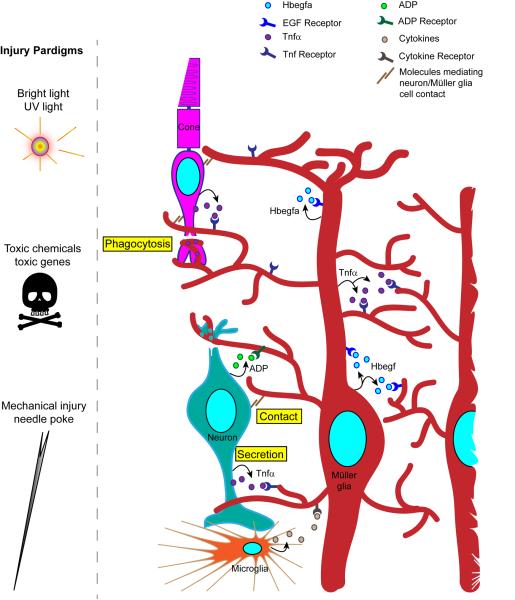
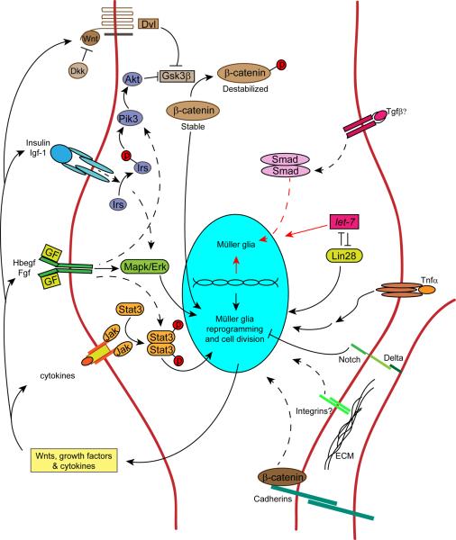
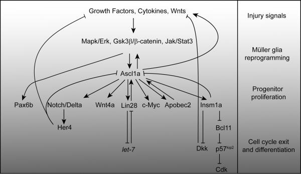
Müller glia are the major glial component of the retina. They are one of the last retinal cell types to be born during development, and they function to maintain retinal homeostasis and integrity. In mammals, Müller glia respond to retinal injury in various ways that can be either protective or detrimental to retinal function. Although these cells can be coaxed to proliferate and generate neurons under special circumstances, these responses are meagre and insufficient for repairing a damaged retina. By contrast, in teleost fish (such as zebrafish), the response of Müller glia to retinal injury involves a reprogramming event that imparts retinal stem cell characteristics and enables them to produce a proliferating population of progenitors that can regenerate all major retinal cell types and restore vision. Recent studies have revealed several important mechanisms underlying Müller glial cell reprogramming and retina regeneration in fish that may lead to new strategies for stimulating retina regeneration in mammals.


Figures

Figure 1
Figure 1. Retinal anatomy
Illustration of the major retinal cell types and their organization in the retina. The retina is divided into 3 laminar layers; the outer nuclear layer (ONL), inner nuclear layer (INL) and ganglion cell layer (GCL). Six different neuronal cell types and one glia cell type are distributed among these layers with rod and cone photorecptors in the ONL; bipolar, horizontal and amacrine interneurons, along with the Müller glia cell bodies, in the INL; and ganglion cells in the GCL. Ganglion cell axons run just beneath the GCL and comprise a nerve fiber layer (NFL). Synapses between photoreceptors and interneurons take place in the outer plexiform layer (OPL) and synapses between interneurons and ganglion cells take place in the inner plexiform layer (IPL). MG processes span all retinal layers and contribute to the formation of the inner limiting membrane (ILM) and outer limiting membrane (OLM). The retinal pigment epithelium (RPE) consists of pigmented cells that absorb light and make contact with photoreceptors.
Figure 2
Figure 2. Generation of multipotent Müller glia-derived progenitors for retinal repair
Adult Müller glia in zebrafish respond to retinal injury by reprogramming their genome (illustrated by a change in the colour of the cell) so that they can acquire stem cell properties,. This reprogramming results in interkinetic nuclear migration to the outer nuclear layer and an asymmetric cell division near the outer limiting membrane. This asymmetric cell division generates a multipotent progenitor that transiently proliferates and restores the original Müller glia. Multipotent progenitors migrate to all cell layers, exit the cell cycle and regenerate all major retinal cell types,.
Figure 3
Figure 3. Injury paradigms and the communication of injury to Müller glia
Various injury paradigms have been used to induce retinal damage and stimulate regeneration in zebrafish. These include prolonged exposure to intense bright light, short exposure to ultraviolet (UV) light; intravitreal injection of toxins (such as oubain and N-methyl-D-aspartate (NMDA)); expression of a toxic gene (such as bacterial nitroreductase, which, in combination with a prodrug generates a cytotoxic product); and mechanical injury (such as that resulting from a needle poke),,,,. Light-based damage paradigms generally destroy a population of photoreceptors, whereas toxins can cause wide-spread damage. Cytotoxic gene products can be directed to specific retinal cell types using appropriate promoters to drive their expression. Mechanical injury generally destroys all retinal cell types in a circumscribed region of the retina. The figure illustrates the ways in which injured cells might communicate with Müller glia to stimulate their reprogramming. These include secretion of signaling molecules (arrows) from damaged cells, Müller glia or infiltrating microglia; altered contact between damaged cells and Müller glia; and phagocytosis of injured cells by Müller glia. Recent studies have suggested that growth factors, such as heparin-binding epidermal growth factor (Hbegf), and cytokines, such as tumor necrosis factor- α (Tnfα), are necessary for Müller glia reprogramming and progenitor formation in the injured retina,. These factors are produced in Müller glia at the injury site and therefore, may act in an autocrine and paracrine fashion. Tnfα and ADP are also released from injured retinal neurons,.
Figure 4
Figure 4. Signaling cascades contributing to Müller glia reprogramming and progenitor proliferation in zebrafish
Retina regeneration requires the activation of a variety of signaling cascades. This diversity of signaling may reflect the variety of injuries and signaling molecules that stimulate retina regeneration. Signaling pathways that have been shown to regulate retina regeneration are indicated by solid arrows, while those indirectly implicated or hypothesized to be involved are indicated by dashed arrows. Secreted factors that regulate Müller glia proliferation are indicated outside the cell (those impacting Müller glia proliferation in birds/mammals, but not yet tested in zebrafish are annotated with a question mark). Arrows pointing to the top half of the nucleus represent pathways that stimulate/maintain Müller glia differentiation/quiescence, while arrows pointing to the bottom half of the nucleus represent pathways that impact Müller glia reprogramming and proliferation. Wnts are secreted lipid-modified glycoproteins that bind Frizzled family receptors to regulate β-catenin stabilization. Dkk (Dickkopf) is a secreted Wnt signaling antagonist. Dvl (Dishevled) is a cytoplasmic phosphoprotein acting downstream of Wnt receptors. Gsk3β (glycogen synthase kinase 3β) regulates β-catenin stabilization by phosphorylation. β-catenin regulates cell adhesion and gene expression. Insulin and Igf-1 (insulin-like growth factor 1) are secreted proteins that bind tyrosine kinase receptors that signal via Irs (insulin receptor signaling protein), an adapter protein that couples insulin and Igf-1 receptor to PI3K (phosphoinositide 3-kinase) and Akt (protein kinase B) activation. Hbegf (heparin binding epidermal-like growth factor) is a transmembrane protein that undergoes ectodomain shedding. It is a member of the Egf family of growth factor ligands and acts via epidermal growth factor receptors. Fgfs (Fibroblast growth factors) are secreted growth factors that bind to fibroblast growth factor receptors. Egf and Fgf receptors are tyrosine kinase receptors that signal via Mapk (mitogen activated protein kinase) and Erk (extracellular signal regulated kinase). Cytokines are secreted proteins that often signal through receptors that lack intrinsic tyrosine kinase activity. Cytokine receptors are often coupled to Jak (Janus kinase) activation. Jak proteins are non-receptor tyrosine kinases that transduce cytokine-mediated signals by phosphorylating Stat proteins (signal transducers and activators of transcription). Tgfβ (Transforming growth factor-beta) is a secreted protein that signals via the Smad pathway to alter gene expression. let-7 is a microRNA that is a posttranscriptional regulator of RNA expression. Lin28 is a RNA binding protein that regulates let-7 microRNA expression. Tnfα (tumor necrosis factor alpha) is a secreted cytokine that acts via TNF receptors to regulate cell signaling and gene expression. Delta-Notch signaling is mediated by single pass trans-membrane proteins expressed on adjacent cells. ECM (extracellular matrix) can signal via transmembrane integrin receptors to regulate cell function.
Figure 5
Figure 5. Regeneration-associated transcriptional cascades in zebrafish
Growth factors, Wnts and cytokines appear to impinge on Mapk/Erk, Gsk3β/β-catenin and Jak/Stat signaling pathways to stimulate Müller glia reprogramming in response to retinal injury,,,. These pathways participate in injury-dependent ascl1a gene expression,,. Ascl1a is a bHLH (basic helix-loop-helix) transcription factor impinging on almost all aspects of retina regeneration. It regulates genes responsible for generating Müller glia-derived progenitors, such as those encoding Wnts, growth factors, Lin28, c-Myc, Apobec2b, Insm1a and Stat3,,,,,,. Ascl1a also controls the expression of proteins and microRNAs that inhibit progenitor formation and proliferation, such as Notch, Dkk, Insm1a (this protein contributes to both progenitor formation and differentiation), p57kip2 and let-7,,,. Gsk3β inhibition stimulates pax6b expression in an Ascl1a-independent fashion. The regenerative steps outlined on the right-hand side, along with the gray gradient, illustrate the gradual transition of Müller glia to progenitors and their differentiation during retina regeneration.

References

    1. Shepherd RK, Shivdasani MN, Nayagam DA, Williams CE, Blamey PJ. Visual prostheses for the blind. Trends Biotechnol. 2013;31:562–571. - PubMed
    1. Pearson RA, et al. Restoration of vision after transplantation of photoreceptors. Nature. 2012;485:99–103. - PMC - PubMed
    2. The first demonstration of vision recovery following rod precursor transplants into a genetic model of blindness.

    1. Boye SE, Boye SL, Lewin AS, Hauswirth WW. A comprehensive review of retinal gene therapy. Mol Ther. 2013;21:509–519. - PMC - PubMed
    1. Sherpa T, et al. Ganglion cell regeneration following whole-retina destruction in zebrafish. Dev Neurobiol. 2008;68:166–181. - PMC - PubMed
    1. Lindsey AE, Powers MK. Visual behavior of adult goldfish with regenerating retina. Vis Neurosci. 2007;24:247–255. - PubMed

Publication types