. 2014 Mar-Apr;34(2):107-14.
doi: 10.5144/0256-4947.2014.107.

Clinical, neuroimaging, and genetic features of L-2-hydroxyglutaric aciduria in Arab kindreds

Affiliations

Clinical, neuroimaging, and genetic features of L-2-hydroxyglutaric aciduria in Arab kindreds

Muhammad Faiyaz-Ul-Haque et al. Ann Saudi Med. 2014 Mar-Apr.

Abstract

Background and objectives: L-2-hydroxyglutaric aciduria is a neurometabolic disorder with autosomal recessive mode of inheritance in which patients exhibit elevated L-2-hydroxyglutaric acid in body fluids, central nervous system manifestations, and increased risk of brain tumor formation. Mutations in L2HGDH gene have been described in L-2-hydroxyglutaric aciduria patients of different ethnicities. The present study was conducted to perform a detailed clinical, imaging and genetic analysis.

Design and settings: A cross-sectional clinical genetic study of 16 L-2-hydroxyglutaric aciduria patients from 4 Arab consanguineous families examined at the metabolic clinic of the hospital.

Patients and methods: Genomic DNA was isolated from the blood of 12 patients and 10 unaffected family members, and the L2HGDH gene was sequenced. DNA sequences were compared to the L2HGDH reference sequence from GenBank.

Results: All patients exhibit characteristic clinical, biochemical, and imaging features of L-2-hydroxyglutaric aciduria, and 4 patients exhibited increased incidence of brain tumors. The sequencing of the L2HGDH gene revealed the c.1015delA, c.1319C > A, and c.169G > A mutations in these patients. These mutations encode for the p.Arg339AspfsX351, p.Ser440Tyr, and p.Gly57Arg changes in the L2HGDH protein, respectively. The c.169G > A mutation, which was shown to have a common origin in Italian and Portuguese patients, was also discovered in Arab patients. Finding of the homozygous c.159T SNP associated with the c.169G > A mutation in Arab patients points to an independent origin of this mutation in Arab population.

Conclusion: The detailed description of clinical manifestations and L2HGDH mutation in this study is useful for diagnosis of L-2-hydroxyglutaric aciduria in Arab patients. While reoccurrence of an L2HGDH mutation in L-2-hydroxyglutaric aciduria patients of different ethnicity is extremely rare, the c.169G mutation has an independent origin in Arab patients. It is likely that this mutation may also be present in patients of other ethnicities.


Figures

Figure 1
Figure 1
Family pedigrees of the L-2-hydroxyglutaric aciduria patients. Patients and unaffected family members whose DNA was sequenced are marked with horizontal lines above their symbols. Double lines indicate consanguineous marriage.
Figure 2
Figure 2
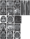
Magnetic resonance imaging (MRI) brain, computed tomography (CT) scan, and magnetic resonance spectroscopy (MRS) images of the patients. MRI, T1- weighted images show high signal intensity involving the subcortical white matter bilaterally (Panels a and b). This mainly involves the peripheral areas of the centrum semiovale and the subcortical new fiber. The similar pattern of patchy increased signal intensity is present on the temporal lobe. The central part of the centrum semiovale in the periventricular region and thalami are preserved. Gray and white matter of the cerebellum is also preserved. The dentate nuclei are swollen and show flow signal intensity on T1-weighted images. T1-weighted sagittal image of the thoracocervical spine showing isointense signal area (arrow) with the surrounding high-intensity rim in the upper cervical cord (Panel c). This lesion (arrow) is hyperintense in T2- weighted image (Panel d). axial T2 image shows the same high signal intensity lesions in the subcortical white matter bilaterally (Panel e). Brain MRI shows prominent dentate nuclei and globi palladi with abnormally increased signal intensity, sparing of the thalami, and extensive leukodystrophy with peripheral predominance (Panels f–i). CT of the brain shows extensive white matter, low-attenuation areas involving both cerebral hemispheres, which is suggestive of leukodystrophy (Panel j). This involves the frontal pole more than the occipital and temporal lobes. MRS shows prominent reduction of the choline peak with mildly decreased N-acetylaspartate peak (Panel k). Coronal MRI of T1-weighted image shows bilateral subcortical white matter changes (*) with sparing of periventricular white matter (+) (Panel l). Sagittal T1-weighted image of brain shows cerebellar dentate nucleus lesion (arrow) (Panel m). Coronal T2-weighted image of brain shows a bilateral dentate nuclear lesion, which is prominent on the left side (arrow) (Panel n). Axial, T2-weighted image of brain shows deep periventricular white matter sparing (+) with the involvement of subcortical white matter (*) (Panel o). The axial T2- weighted image of brain shows sparing of thalami (+) with the involvement of caudate (x) and lentiform nuclei (arrow) with subcortical white matter changes (*) (Panel p). CT of brain axial image shows subcortical white matter changes bilaterally (arrows) (P and q).
Figure 3
Figure 3
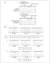
(A) Chromatogram of a patient with the c.169G>A mutation. The upper panel depicts the homozygous c.169G>A mutation and the homozygous c.159T single-nucleotide polymorphism (SNP) in the L2HGDH gene of a patient from family 4. The lower panel shows the wild-type homozygous c.169G and the homozygous c.159T SNP in the L2HGDH gene of an unaffected individual from family 4. (B) Sequence analyses of mutations in the L2HGDH gene. Homozygous mutations in L2H aciduria patients are shown in sequence chromatograms on the left (arrowheads), heterozygous (carrier) sequence chromatograms on the middle, and normal sequences from control subjects on the right panels. The upper panel (i) depicts the p.Arg339AspfsX351 mutation from family 1 and family 3, the middle panel (ii) depicts the p.Ser440Tyr mutation from family 2, and the lower panel (iii) shows the p.Gly57Arg mutation from family 4.

Similar articles

Cited by

References

    1. Duran M, Kamerling JP, Bakker HD, van Gennip AH, Wadman SK. L-2-Hydroxyglutaric aciduria: an inborn error of metabolism? J Inherit Metab Dis. 1980;3(4):109–112. - PubMed
    1. Steenweg ME, Jakobs C, Errami A, van Dooren SJ, Adeva Bartolomé MT, et al. An overview of L-2-hydroxyglutarate dehydrogenase gene (L2H-GDH) variants: a genotype-phenotype study. Hum Mutat. 2010;31(4):380–390. Review. - PubMed
    1. Steenweg ME, Salomons GS, Yapici Z, Uziel G, Scalais E, Zafeiriou DI, et al. L-2-Hydroxyglutaric aciduria: pattern of MR imaging abnormalities in 56 patients. Radiology. 2009;251(3):856–865. - PubMed
    1. Haliloglu G, Jobard F, Oguz KK, Anlar B, Akalan N, Coskun T, et al. L-2-hydroxyglutaric aciduria and brain tumors in children with mutations in the L2HGDH gene: neuroimaging findings. Neuropediatrics. 2008;39(2):119–122. - PubMed
    1. Moroni I, Bugiani M, D’Incerti L, Maccagnano C, Rimoldi M, et al. L-2-hydroxyglutaric aciduria and brain malignant tumors: a predisposing condition? Neurology. 2004;62(10):1882–1884. Review. - PubMed

Publication types

MeSH terms

Substances

Supplementary concepts