. 2014 Jun 27;77(6):1473-87.
doi: 10.1021/np5002384. Epub 2014 Jun 4.

Essential parameters for structural analysis and dereplication by (1)H NMR spectroscopy

Affiliations

Essential parameters for structural analysis and dereplication by (1)H NMR spectroscopy

Guido F Pauli et al. J Nat Prod. .

Abstract

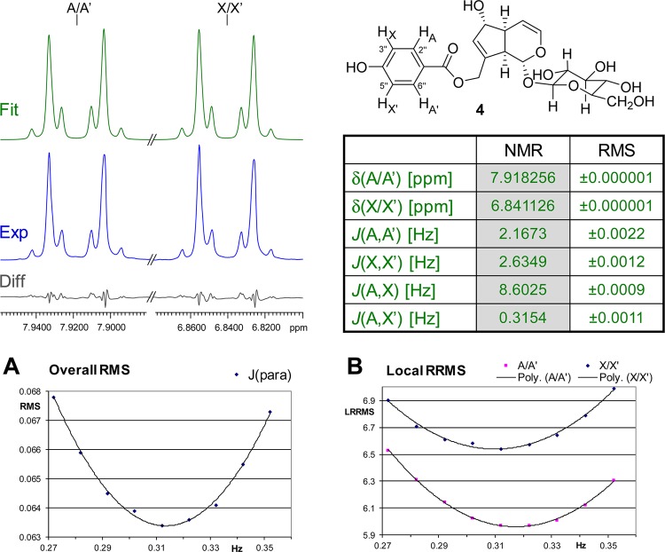
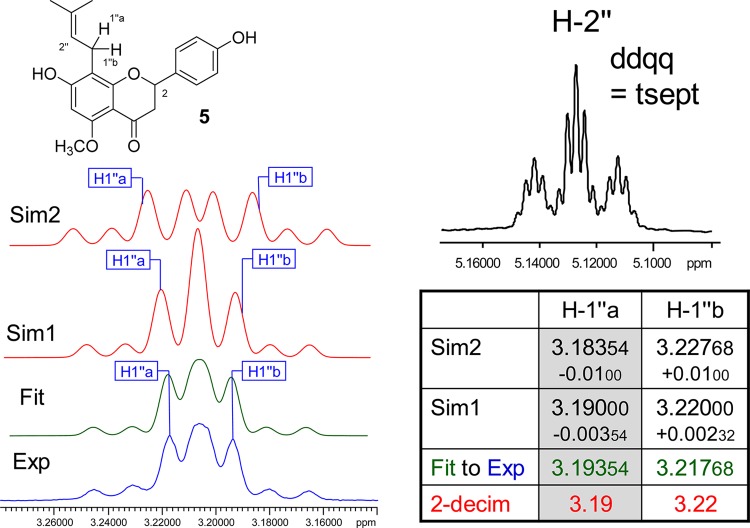
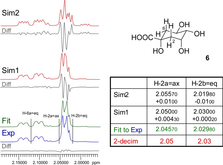
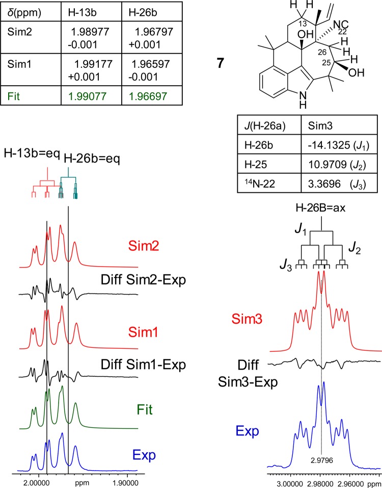
The present study demonstrates the importance of adequate precision when reporting the δ and J parameters of frequency domain (1)H NMR (HNMR) data. Using a variety of structural classes (terpenoids, phenolics, alkaloids) from different taxa (plants, cyanobacteria), this study develops rationales that explain the importance of enhanced precision in NMR spectroscopic analysis and rationalizes the need for reporting Δδ and ΔJ values at the 0.1-1 ppb and 10 mHz level, respectively. Spectral simulations paired with iteration are shown to be essential tools for complete spectral interpretation, adequate precision, and unambiguous HNMR-driven dereplication and metabolomic analysis. The broader applicability of the recommendation relates to the physicochemical properties of hydrogen ((1)H) and its ubiquity in organic molecules, making HNMR spectra an integral component of structure elucidation and verification. Regardless of origin or molecular weight, the HNMR spectrum of a compound can be very complex and encode a wealth of structural information that is often obscured by limited spectral dispersion and the occurrence of higher order effects. This altogether limits spectral interpretation, confines decoding of the underlying spin parameters, and explains the major challenge associated with the translation of HNMR spectra into tabulated information. On the other hand, the reproducibility of the spectral data set of any (new) chemical entity is essential for its structure elucidation and subsequent dereplication. Handling and documenting HNMR data with adequate precision is critical for establishing unequivocal links between chemical structure, analytical data, metabolomes, and biological activity. Using the full potential of HNMR spectra will facilitate the general reproducibility for future studies of bioactive chemicals, especially of compounds obtained from the diversity of terrestrial and marine organisms.


Figures

Chart 1
Chart 1
Figure 1
Figure 1
Case study 1: uzarigenin-3-sulfate (1). Representing the class of steroidal natural products, 1 belongs to the 5α series and, thus, is a case of rather disperse δ distribution of the steroidal envelope. Given are the experimental spectrum (Exp, in blue) with accurate assignments of the nine protons in the region, compared with the simulated spectrum with chemical shifts rounded to 0.01 ppm for the methylene protons, H-11A/B and H-12A/B only (Sim, in red). The difference spectrum (Diff, in gray) clarifies the considerable deviations caused by the inappropriate rounding of δ values to two decimals, which translates into a visual mismatch of the “fingerprint” region of steroid HNMR spectra and can invalidate dereplication of these stereochemically demanding natural products (600 MHz, methanol-d4).
Figure 2
Figure 2
Case study 2: progesterone (2). Even at ultrahigh magnetic field, the steroid 2, like many alicyclic terpenoids, exhibits overlapping resonances. HiFSA can produce complete δ/J profiles and fully fitted spectra (Fit, in green). As shown for the overlapping resonances of the two notably uncoupled methylene proton pairs, H-2a/b and H-6a/b (overview A, expansions B and C), reporting with only 0.01 ppm precision produces marked deviations in the resulting simulated spectra (Sim, in red), which in the case of H-2b and H-6a even results in a reversal of chemical shift order and misassignment of the signals (900 MHz, methanol-d4). The difference spectra (Diff, in gray) show the extent of the mismatch produced by such inadequate reporting artifacts.
Figure 3
Figure 3
Case study 3: syringetin-3-O-β-d-glucoside (3). Quantum mechanical simulation of the spin systems (scenarios Sim1–3, in red) shows that minor variations of the chemical shifts of the three protons H-1″, H-2″, and H-3″ of the glucose moiety lead to major deviations from the fitted spectrum (Fit, in green; matching the experimental data, Exp, in blue). All J values were kept constant for the simulations (600 MHz, methanol-d4).
Figure 4
Figure 4
Case study 4: agnuside (4). The complex aromatic resonances of the widely occurring para-substituted phenyl structural motif result from the underlying AA′XX′ (in 2), AA′MM′, or AA′BB′ (in analogous molecules) spin systems. Their precise numerical description was performed using the HiFSA approach and requires δ and J reporting precision to the low ppb and mHz levels, respectively. Shown on the top left are the experimental (Exp, in blue) and HiFSA fitted (Fit, in green) spectra, their residual difference (Diff, in gray), and the tabulated spin parameters and RMS values of the fit (360 MHz, methanol-d4). Panel A: Plot of the overall residual RMS for different J values for the para-coupling 5JA,X′. Panel B: Plot of the local RRMSs for A/A′ and X/X′ with different J values for the para-coupling 5JA,X′.
Figure 5
Figure 5
Case study 5: isoxanthohumol (5). Owing to the influence of the C-2 stereogenic center and the aromatic ring isotropy, the methylene protons H2-1″ of 5 are diastereotopic. Accordingly, there are two resonances, H-1″a and H-1″b, which exhibit a small but important chemical shift difference (Δδ). While HiFSA fitting (Fit, in green) yields the precise spectral parameters, even small deviations of only the δ values lead to major changes in the simulated spectra and, thus, would impede dereplication. All J values were kept constant for the simulations (Sim, in red). A notable detail of the prenyl motif is the highly characteristic resonance of the olefinic proton, H-2″, which is coupled with all other protons in the prenyl moiety (500.163 MHz, methanol-d4).
Figure 6
Figure 6
Case study 6: quinic acid (6). Quinic acid derivatives, such as chlorogenic acid, formed by esterification with cinnamates occur widely in the plant kingdom and exhibit various degrees of diastereotopism of the C-2 methylene protons. As shown here for the core molecule, 6, the chirality induces a small but important chemical shift difference for H-2a vs H-2b. Only precise HiFSA fitting yields a congruent spectrum (Fit, in green), whereas even very small misalignments in the low ppb and even ppt range such as in Sim1 (Δδ of H-2a = 200 ppt, H-2b = 4.3 ppb) lead to mismatching of the resulting simulated spectra (360 MHz, methanol-d4).
Figure 7
Figure 7
Case study 7: ambiguine N isonitrile (7). This case shows that high precision is required to properly document the resonances of two apparently “un(cor)related” protons in a molecule: although the two closely resonating protons, H-13b and H-26b, are not coupled to each other, perturbations as low as ±0.001 ppm (Sim1 + 2, in red) still have striking effects on the spectra compared to the experimental spectrum (Exp, in blue). A special feature of 7 is the heteronuclear 3J-coupling of H-26a with N-22, which is a rarely described property that actually can be used to distinguish molecules within alkaloid classes, such as the ambiguines: The spin-1 nucleus, 14N, gives rise to a triplet coupling pattern with a specific 1:1:1 line intensity (Sim3, in red). All J values were kept constant for the simulation (900 MHz, methanol-d4).

References

    1. Reynolds W. F.; Enriquez R. G. J. Nat. Prod. 2002, 65, 221–244. - PubMed
    1. Claridge T. D. W.High-Resolution NMR Techniques in Organic Chemistry, 1st ed.; Pergamon: Amsterdam, 1999.
    1. Chamberlain N. F.The Practice of NMR Spectroscopy with Spectra-Structure Correlations for Hydrogen-1; Plenum Press: New York, 1974.
    1. Nicolaou K. C.; Snyder S. A. Angew. Chem., Int. Ed. 2005, 44, 1012–1044. - PubMed
    1. Robien W. Trends Anal. Chem. 2009, 28, 914–922.

Publication types

LinkOut - more resources