. 2014 Jun 3;3(3):e000716.
doi: 10.1161/JAHA.113.000716.

Protein changes contributing to right ventricular cardiomyocyte diastolic dysfunction in pulmonary arterial hypertension

Affiliations

Protein changes contributing to right ventricular cardiomyocyte diastolic dysfunction in pulmonary arterial hypertension

Silvia Rain et al. J Am Heart Assoc. .

Abstract

Background: Right ventricular (RV) diastolic function is impaired in patients with pulmonary arterial hypertension (PAH). Our previous study showed that elevated cardiomyocyte stiffness and myofilament Ca(2+) sensitivity underlie diastolic dysfunction in PAH. This study investigates protein modifications contributing to cellular diastolic dysfunction in PAH.

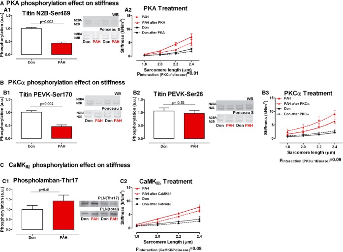
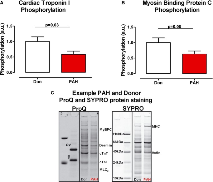
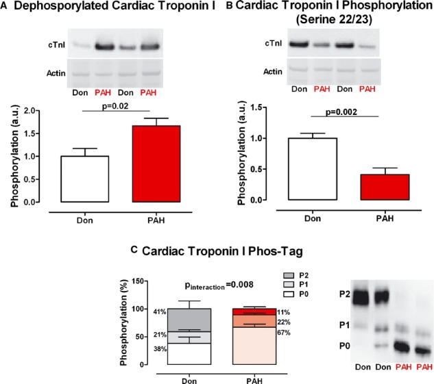
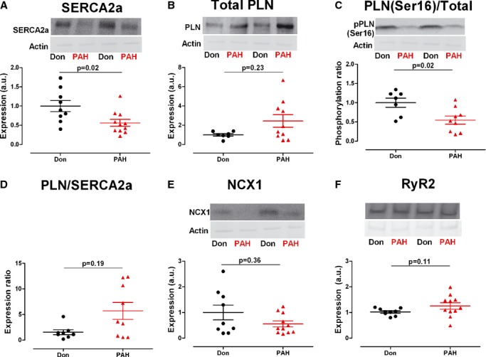
Methods and results: RV samples from PAH patients undergoing heart-lung transplantation were compared to non-failing donors (Don). Titin stiffness contribution to RV diastolic dysfunction was determined by Western-blot analyses using antibodies to protein-kinase-A (PKA), Cα (PKCα) and Ca(2+)/calmoduling-dependent-kinase (CamKIIδ) titin and phospholamban (PLN) phosphorylation sites: N2B (Ser469), PEVK (Ser170 and Ser26), and PLN (Thr17), respectively. PKA and PKCα sites were significantly less phosphorylated in PAH compared with donors (P<0.0001). To test the functional relevance of PKA-, PKCα-, and CamKIIδ-mediated titin phosphorylation, we measured the stiffness of single RV cardiomyocytes before and after kinase incubation. PKA significantly decreased PAH RV cardiomyocyte diastolic stiffness, PKCα further increased stiffness while CamKIIδ had no major effect. CamKIIδ activation was determined indirectly by measuring PLN Thr17phosphorylation level. No significant changes were found between the groups. Myofilament Ca(2+) sensitivity is mediated by sarcomeric troponin I (cTnI) phosphorylation. We observed increased unphosphorylated cTnI in PAH compared with donors (P<0.05) and reduced PKA-mediated cTnI phosphorylation (Ser22/23) (P<0.001). Finally, alterations in Ca(2+)-handling proteins contribute to RV diastolic dysfunction due to insufficient diastolic Ca(2+) clearance. PAH SERCA2a levels and PLN phosphorylation were significantly reduced compared with donors (P<0.05).

Conclusions: Increased titin stiffness, reduced cTnI phosphorylation, and altered levels of phosphorylation of Ca(2+) handling proteins contribute to RV diastolic dysfunction in PAH.

Keywords: diastole; pulmonary heart disease.


Figures

Figure 1.
Figure 1.
PKA, PKCα, and CaMKIIδ treatment effect on diastolic stiffness mediated by titin phosphorylation. A‐A1. Titin N2B domain serine 469 PKΑ‐dependent phosphorylation. Typical example of the 2 titin isoforms (upper band: N2BA; lower band N2B) immunostained with phosphospecific antibody against serine 469 site on titin N2B domain and the corresponding Ponceau S staining for total titin (nDon=7, nPAH=5). A2. Donor and PAH cardiomyocyte stiffness was measured in relaxing solution at increasing sarcomere length (1.8 to 2.4 μm)—continuous line. The same cardiomyocyte was further incubated with PKA active subunit and stiffness measurements were repeated—dotted line (nDon=4, nPAH=4). B‐B1. Titin PEVK domain serine 170 PKCα‐dependent phosphorylation. Typical example of the 2 titin isoforms (upper band: N2BA; lower band N2B) immunostained with phosphospecific antibody against serine 170 site on titin PEVK domain and the corresponding Ponceau S staining for total titin (nDon=7, nPAH=5). B2.Titin PEVK domain serine 26 PKCα‐dependent phosphorylation. Typical example of the 2 titin isoforms (upper band: N2BA; lower band N2B) immunostained with phosphospecific antibody against serine 26 site on titin PEVK domain and the corresponding Ponceau S staining for total titin (nDon=7, nPAH=5). B3. Donor and PAH cardiomyocyte stiffness was measured in relaxing solution at increasing sarcomere length (1.8 to 2.4 μm)—continuous line. The same cardiomyocyte was further incubated with PKCα active subunit and stiffness measurements were repeated—dotted line (nDon=3, nPAH=3). C‐C1. Phospholamban threonine 17 CamKIIδ‐dependent phosphorylation was used as an indirect measurement of titin CamKIIδ phosphorylation. The phosphorylation level was normalized to the total amount of Phospholamban present in the sample (nDon=7, nPAH=11). C2. Donor and PAH cardiomyocyte stiffness was measured in relaxing solution at increasing sarcomere length (1.8 to 2.4 μm)—continuous line. The same cardiomyocyte was further incubated with CamKIIδ and stiffness measurements were repeated—dotted line (nDon=3, nPAH=3). Data presented as mean±SEM. CamKIIδ indicates calmoduling‐dependent‐kinase; PAH, pulmonary arterial hypertension; PKA, protein‐kinase‐A; PLN, phospholamban.
Figure 2.
Figure 2.
Sarcomeric protein phosphorylation. A, cTnI total phosphorylation was determined by ProQ‐Diamond staining, while total protein content was determined by SYPRO‐Ruby staining (nDon=10, nPAH=10). B, MyBPC total phosphorylation was determined by ProQ staining, while total protein content was determined by SYPRO staining (nDon=10, nPAH=10). C, Typical example of donor and PAH samples ProQ and SYPRO staining and the relative location of specific sarcomeric protein. ProQ staining annotations—ov, ovalbumin; β, β‐casein; MyBPC, myosin binding protein C; cTnT, cardiac troponin T; cTnI, cardiac troponin I; MLC2, myosin light chain 2. SYPRO staining annotations—MHC, myosin heavy chain. Data presented as mean±SEM. PAH inidcates pulmonary arterial hypertension.
Figure 3.
Figure 3.
cTnI phosphorylation. A, Typical example of donor and PAH samples immunostained against the unphosphorylated form of cTnI and normalized for Ponceau‐S stained actin. cTnI total phosphorylation was determined by Western blot analysis in donor and PAH samples (nDon=8, nPAH=9). B, Typical example of donor and PAH samples immunostained against cTnI serine 22/23 residue and normalized for Ponceau‐S stained actin. cTnI serine 22/23 phosphorylation was determined by Western blot analysis in donor and PAH samples (nDon=9, nPAH=11). C, Typical example of donor and PAH samples immunostained against unphosphorylated (P0) cTnI, mono‐phosphorylated (P1) cTnI and bis‐phosphorylated (P2) cTnI. cTnI relative phosphorylation distribution was determined by Phos‐Tag analysis in donor and PAH samples (nDon=5, nPAH=9). Data presented as mean±SEM. cTnI indicates cardiac troponin I; cTnT, cardiac troponin T; PAH, pulmonary arterial hypertension.
Figure 4.
Figure 4.
Ca2+‐handling proteins expression and phosphorylation. Typical example of donor and PAH samples immunostained against the corresponding Ca2+‐handeling protein and normalized for Ponceau‐S stained actin. A, SERCA2a: Sarco/Endoplasmic Reticulum Ca2+‐ATPase 2a (cardiac isoform) expression level (nDon=9, nPAH=11). B, Total PLN: phospholamban expression level (nDon=7, nPAH=10). C, PLN (Ser16)/Total PKA‐dependent phosphorylation level of serine 16 residue on PLN normalized to total PLN protein content (nDon=7, nPAH=9). D, PLN/SERCA2a inhibitory effect of PLN on SERCA2a quantified by the ratio between 2 (nDon=8, nPAH=9). E, NCX1, Na+/Ca2+ exchanger 1 (cardiac isoform) expression level (nDon=9, nPAH=11). F, RyR2, ryanodine receptor 2 (cardiac isoform) expression level (nDon=9, nPAH=11). Data presented as mean±SEM. PAH indicates pulmonary arterial hypertension; PLA, protein‐kinase‐A; PLN, phospholamban.

References

    1. Rain S, Handoko ML, Trip P, Gan CT‐J, Westerhof N, Stienen GJ, Paulus WJ, Ottenheijm CAC, Marcus JT, Dorfmüller P, Guignabert C, Humbert M, Macdonald P, Dos Remedios C, Postmus PE, Saripalli C, Hidalgo CG, Granzier HL, Vonk‐Noordegraaf A, van der Velden J, de Man FS. Right ventricular diastolic impairment in patients with pulmonary arterial hypertension. Circulation. 2013; 128:2016-2025. - PubMed
    1. De Tombe PP, Granzier HL. The cytoskeleton and the cellular transduction of mechanical strain in the heart: a special issue. Pflugers Arch. 2011; 462:1-2. - PubMed
    1. Fukuda N, Granzier HL, Ishiwata S, Kurihara S. Physiological functions of the giant elastic protein titin in mammalian striated muscle. J Physiol Sci. 2008; 58:151-159. - PubMed
    1. Li S, Guo W, Schmitt BM, Greaser ML. Comprehensive analysis of titin protein isoform and alternative splicing in normal and mutant rats. J Cell Biochem. 2012; 113:1265-1273. - PMC - PubMed
    1. Hidalgo C, Hudson B, Bogomolovas J, Zhu Y, Anderson B, Greaser M, Labeit S, Granzier H. PKCΑ phosphorylation of titin's PEVK element: a novel and conserved pathway for modulating myocardial stiffness. Circ Res. 2009; 105:631-638. - PMC - PubMed

Publication types

MeSH terms

Substances