Case Reports
. 2014 Jul;76(1):140-7.
doi: 10.1002/ana.24192. Epub 2014 Jun 20.

A fatal case of JC virus meningitis presenting with hydrocephalus in a human immunodeficiency virus-seronegative patient

Affiliations
Case Reports

A fatal case of JC virus meningitis presenting with hydrocephalus in a human immunodeficiency virus-seronegative patient

Shruti P Agnihotri et al. Ann Neurol. 2014 Jul.

Abstract

JC virus (JCV) is the etiologic agent of progressive multifocal leukoencephalopathy, JCV granule cell neuronopathy, and JCV encephalopathy. Whether JCV can also cause meningitis has not yet been demonstrated. We report a case of aseptic meningitis resulting in symptomatic hydrocephalus in a human immunodeficiency virus-seronegative patient. Brain imaging showed enlargement of ventricles but no parenchymal lesion. She had a very high JC viral load in the cerebrospinal fluid (CSF) and developed progressive cognitive dysfunction despite ventricular drainage. She was diagnosed with pancytopenia and passed away after 5.5 months. Postmortem examination revealed productive JCV infection of leptomeningeal and choroid plexus cells, and limited parenchymal involvement. Sequencing of JCV CSF strain showed an archetype-like regulatory region. Further studies of the role of JCV in aseptic meningitis and in idiopathic hydrocephalus are warranted.


Figures

Figure 1
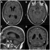
Figure 1. Magnetic Resonance Imaging of the brain demonstrates hydrocephalus and abnormal signal in subarachnoid space
MRI performed 2.5 months after symptoms onset shows hydrocephalus and no parenchymal lesions. Panels A and B are axial FLAIR sequences showing enlarged ventricles and abnormal hyperintensity in the subarachnoid space, within the sulci of the cerebral hemispheres (arrows). Panel C is a sagittal T1-weighted sequence demonstrating significant enlargement of the lateral ventricle. In Panel D, there was no enhancement of the meninges post intravenous contrast administration, as seen on this coronal section.
Figure 2
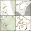
Figure 2. Immunohistochemistry (IHC) analyses of JCV infection in the CNS
Panel A shows double IHC staining for JCV T Ag (brown) and JCV VP1 protein (blue). Numerous JCV-infected leptomeningeal cells expressing VP1 protein (long arrow) or T Ag (short arrow) are overlying the cerebral cortex. A JCV-infected cell can be seen in the subpial gray matter (GM) of the cerebral cortex, expressing T Ag (arrowhead) Bar: 25um. Panel B shows a lower magnification of the cerebral cortex, which reveals many JCV-infected cells in the leptomeninges (long arrows) as well as in the walls of meningeal vessels (short arrows), and few JCV infected cells in the cortical GM (arrowheads). Bar: 200um. Panel C shows a high magnification of the cerebellum (boxed area in the inset), with a subpial cell in the molecular layer (ML) expressing JCV VP1 protein (arrowhead) close to projecting fibers of cells from the granule cell layer (GCL) expressing JCV T Ag (long arrows). Isolated JCV-infected cells, expressing T Ag can be seen in the GCL (short arrows). Bar: 50um. Panel D shows JCV-infected epithelial cells of the choroid plexus of the lateral ventricles expressing VP1 protein (long arrows) or T Ag (short arrows). Bar: 25um.
Figure 3
Figure 3. Comparison of JCV regulatory regions (RR)
The prototype CNS strain JCVMad-1 has two identical 98 bp elements, whereas the archetype RR, commonly found in kidneys and urine samples has a single 98 bp element and a 23 bp and 66 bp inserts. The JCVM1 RR from the CSF of the JCV meningitis patient has an archetype-like RR with two single nucleotide mutations in the 98 bp element and partial deletion of the 66 bp insert. This insert is located at nt position 93, instead of nt position 92 in the archetype RR.

References

    1. Gheuens S, Wuthrich C, Koralnik IJ. Progressive multifocal leukoencephalopathy: why gray and white matter. Annu Rev Pathol. 2013 Jan 24;8:189–215. - PubMed
    1. Behzad-Behbahani A, Klapper PE, Vallely PJ, Cleator GM, Bonington A. BKV-DNA and JCV-DNA in CSF of patients with suspected meningitis or encephalitis. Infection. 2003 Dec;31(6):374–8. - PubMed
    1. Blake K, Pillay D, Knowles W, Brown DW, Griffiths PD, Taylor B. JC virus associated meningoencephalitis in an immunocompetent girl. Arch Dis Child. 1992 Jul;67(7):956–7. - PMC - PubMed
    1. Viallard JF, Ellie E, Lazaro E, Lafon ME, Pellegrin JL. JC virus meningitis in a patient with systemic lupus erythematosus. Lupus. 2005;14(12):964–6. - PubMed
    1. Wuthrich C, Koralnik IJ. Frequent infection of cortical neurons by JC virus in patients with progressive multifocal leukoencephalopathy. J Neuropathol Exp Neurol. 2012 Jan;71(1):54–65. - PMC - PubMed

Publication types