. 2014 Aug;23(8):1113-22.
doi: 10.1002/pro.2497. Epub 2014 Jun 17.

Fine tuning of the catalytic activity of colicin E7 nuclease domain by systematic N-terminal mutations

Affiliations

Fine tuning of the catalytic activity of colicin E7 nuclease domain by systematic N-terminal mutations

Eszter Németh et al. Protein Sci. 2014 Aug.

Abstract

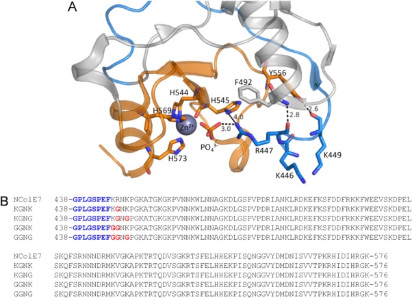
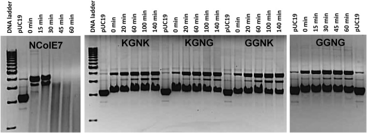
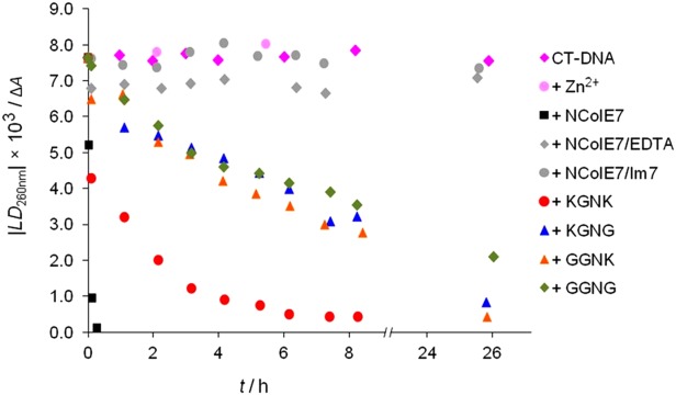
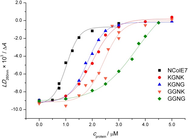
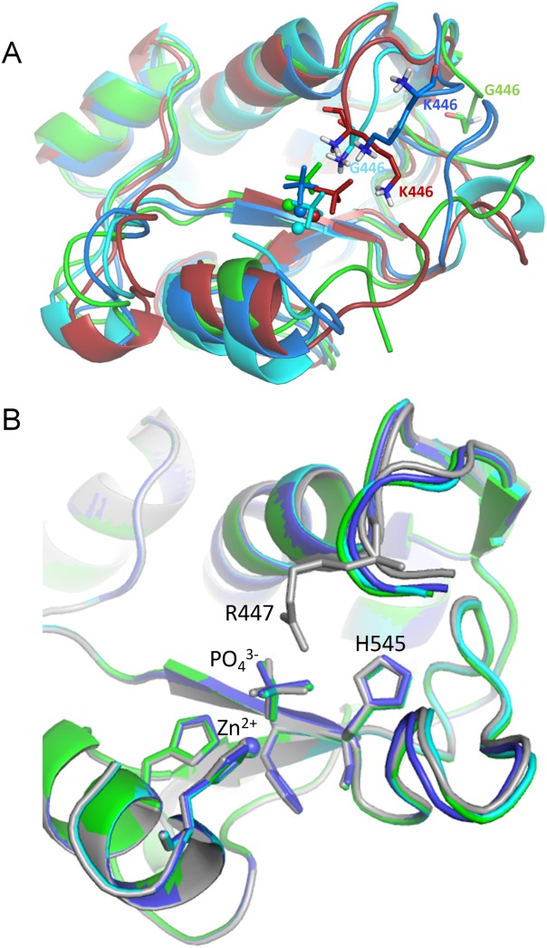
The nuclease domain of colicin E7 (NColE7) promotes the nonspecific cleavage of nucleic acids at its C-terminal HNH motif. Interestingly, the deletion of four N-terminal residues (446-449 NColE7 = KRNK) resulted in complete loss of the enzyme activity. R447A mutation was reported to decrease the nuclease activity, but a detailed analysis of the role of the highly positive and flexible N-terminus is still missing. Here, we present the study of four mutants, with a decreased activity in the following order: NColE7 >> KGNK > KGNG ∼ GGNK > GGNG. At the same time, the folding, the metal-ion, and the DNA-binding affinity were unaffected by the mutations as revealed by linear and circular dichroism spectroscopy, isothermal calorimetric titrations, and gel mobility shift experiments. Semiempirical quantum chemical calculations and molecular dynamics simulations revealed that K446, K449, and/or the N-terminal amino group are able to approach the active centre in the absence of the other positively charged residues. The results suggested a complex role of the N-terminus in the catalytic process that could be exploited in the design of a controlled nuclease.

Keywords: DNA cleavage; Zn2+; binding; flow linear dichroism; isothermal calorimetry; positively charged N-terminal residues.


Figures

Figure 1
Figure 1
(A) Crystal structure of NColE7 (PDB: 1MZ86) in complex with a Zn2+ and a phosphate ion. The HNH motif is in orange and the N-terminal loop is in blue. Among the N-terminal amino acids, R447 is the closest residue to the phosphate ion that forms a bridge between the Zn2+-ion and R447. Hydrogen bonds of the backbone of the N-terminus are also shown. (B) Sequences of the purified NColE7 variants. The proteins containing the black part of the sequence denoted by NColE7 will be referred to as wild type (WT) NColE7 in the following text. The four mutants are named KGNK, KGNG, GGNK, and GGNG based on the 446–449 segments of the sequence, respectively. The changes compared to the NColE7 sequence are highlighted in red. The remaining sequence after the Human rhinovirus C3 protease cleavage encoded by pGEX-6P-1 plasmid is written in blue.
Figure 2
Figure 2
Digestion of 28 nM pUC19 (that is 74 µM calculated for base pairs) by 2.8 µM NColE7 mutants, incubated with one equivalent Zinc(II)-acetate before mixing with DNA. The samples were kept at 37°C and run subsequently on 1% agarose gel. In control experiments (data not shown), cleavage of the plasmid DNA incubated with only Zinc(II)-acetate was not observed. 1 kb Molecular Ruler (BioRad) served as the reference.
Figure 3
Figure 3
Cleavage of 130 µM (base pairs) CT-DNA by different nucleases (0.5 µM) followed by FLD spectroscopy. The proteins were preincubated for 30 min in the presence of one equivalent ZnCl2. The LD signal intensity at 260 nm is taken from baseline corrected spectra of aliquots of the stock solutions, incubated at 37°C. Control experiments included CT-DNA incubated at 37°C without or in the presence of 0.5 µM ZnCl2, 0.5 µM NColE7, and 60 µM EDTA or 0.5 µM NColE7 and 1 µM Im7.
Figure 4
Figure 4
(A) SRCD spectra of the NColE7 variant proteins. (B) SRCD spectra of WT NColE7 in the absence (two independent measurements) and upon the addition of Zn2+-ions. All spectra were normalized to the same 31.6 µM protein concentration.
Figure 5
Figure 5
Microcalorimetric titration of 50 µM KGNG in 20 mM cacodylate buffer, pH = 7.0. The red points at the beginning were ignored during the curve evaluation.
Figure 6
Figure 6
The effect of the CT-DNA binding of NColE7 and the four mutants on the FLD signal intensity at 260 nm. 130 µM CT-DNA (calculated for base pairs) was incubated with 0–5 µM protein and 60 µM EDTA in each case.
Figure 7
Figure 7
(A) Snapshots at 20 ns of the simulations of the mutant proteins: KGNK in red, KGNG in blue, GGNK in cyan, and GGNG in green. The Zn2+-ions are shown by spheres and the phosphate ion by sticks. (B) Optimized structure of the mutants and the WT NColE7. NColE7 is in grey, KGNG in blue, GGNK in cyan, and GGNG in green. Molecules were aligned by PyMOL.

References

    1. Chak K, Kuo W, Lu F, James R. Cloning and characterization of the Cole7 plasmid. J Gen Microbiol. 1991;137:91–100. - PubMed
    1. Lin Y, Liao C, Liang P, Yuan H, Chak K. Involvement of colicin in the limited protection of the colicin producing cells against bacteriophage. Biochem Biophys Res Commun. 2004;318:81–87. - PubMed
    1. Liao C, Hsiao K, Liu Y, Leng P, Yuen HS, Chak K. Processing of DNase domain during translocation of colicin E7 across the membrane of Escherichia coli. Biochem Biophys Res Commun. 2001;284:556–562. - PubMed
    1. Cheng YS, Shi Z, Doudeva LG, Yang WZ, Chak KF, Yuan HS. High-resolution crystal structure of a truncated ColE7 translocation domain: implications for colicin transport across membranes. J Mol Biol. 2006;356:22–31. - PubMed
    1. Shi Z, Chak K, Yuan H. Identification of an essential cleavage site in ColE7 required for import and killing of cells. J Biol Chem. 2005;280:24663–24668. - PubMed

Publication types