

Case Reports
doi: 10.1007/s13139-012-0180-6.
Epub 2012 Oct 15.
Paraneoplastic Cerebellar Degeneration as Initial Presentation of Papillary Carcinoma of the Fallopian Tube: Evaluation and Usefulness of (18)F-FDG PET-CT. Case Report and Literature Review
Affiliations
- PMID: 24895509
- PMCID: PMC4035212
- DOI: 10.1007/s13139-012-0180-6
Item in Clipboard
Case Reports
Paraneoplastic Cerebellar Degeneration as Initial Presentation of Papillary Carcinoma of the Fallopian Tube: Evaluation and Usefulness of (18)F-FDG PET-CT. Case Report and Literature Review
Nucl Med Mol Imaging.
2013 Mar.
Abstract
The acquisition of an (18)F-FDG PET-CT scan in patients with suspected paraneoplastic cerebellar syndrome can be helpful in determining the origin of a neoplasm because of its high sensitivity and also helps guide the neurological development course depending on the degree of incorporation of (18)F-FDG to the cerebellar parenchyma when compared with the rest of the brain.
Figures
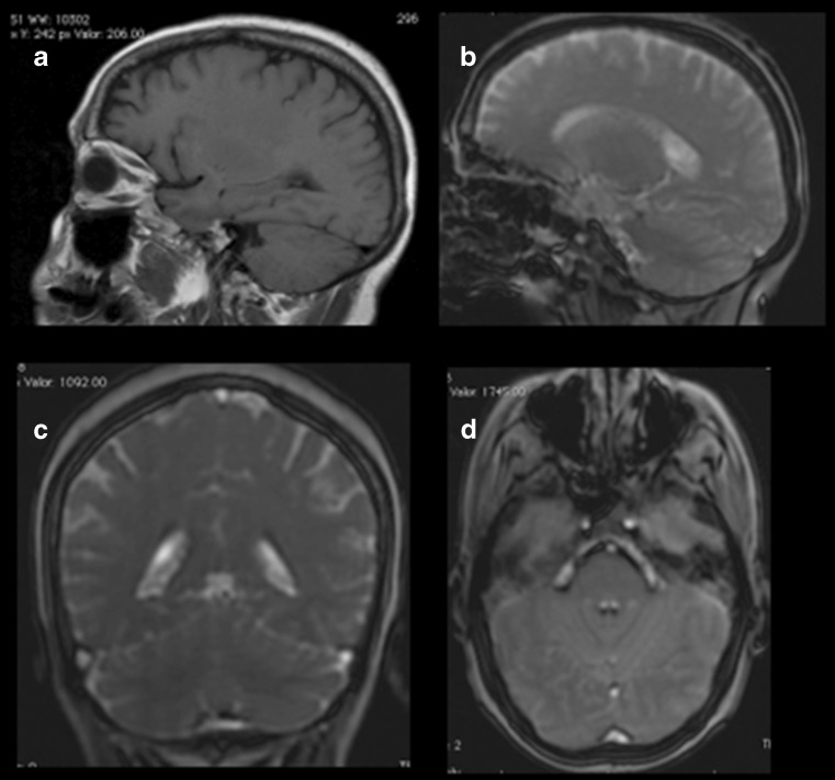
Initial MRI findings were normal. a Non-contrast-enhanced sagittal-slice T1-weighted image. b, c, and d Sagittal, coronal, and trans-axial T2-weighted image, respectively
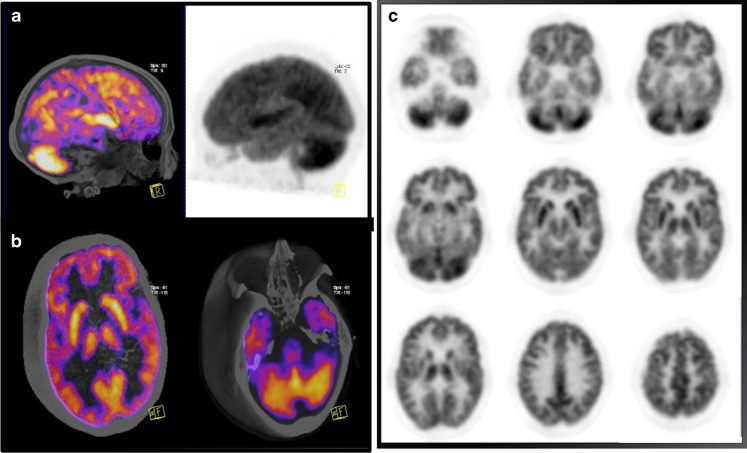
18F-FDG PET-CT brain scan. a 3D fused PET-CT sagittal slice and MIP reconstruction show increased metabolism in both cerebellar hemispheres. b 3D fused PET-CT axial slices. c Whole-brain axial slices
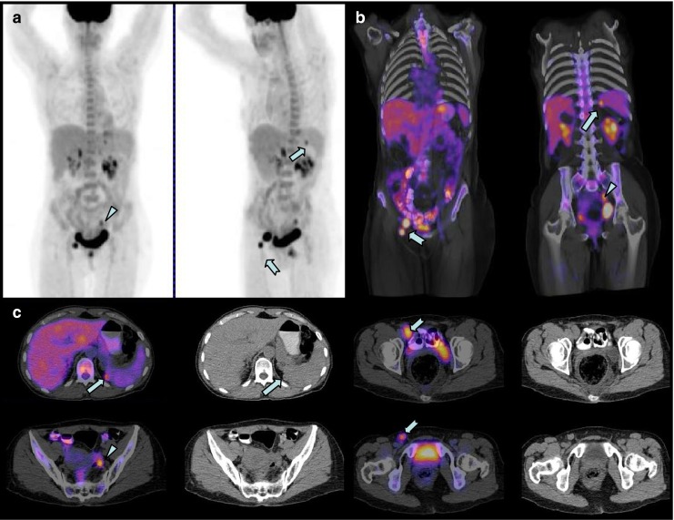
18F-FDG whole body PET-CT. a and b MIP coronal reconstruction showing a focal hypermetabolism in the left pelvis, corresponding to the site of the primary tumor (Fallopian tube topography) (arrowhead). c Right inguinal lymphadenopathy and a spleen focal lesion corresponding to metastatic disease (thick arrow)

Hematoxylin-eosin staining shows edema in the villi of the Fallopian tubes, semi-solid and papillary formation of neoplastic cells. a Normal tissue); b neoplastic tissue
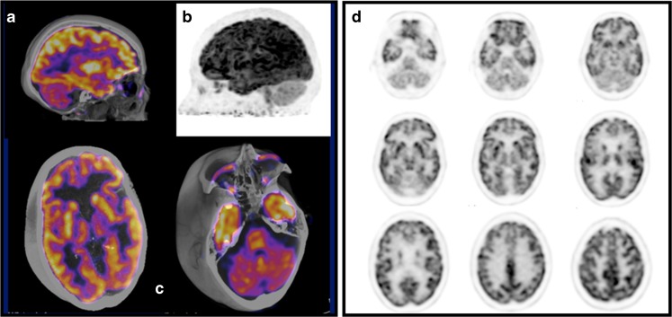
18F-FDG PET-CT brain scan after immunoglobulin and oncological therapy. a 3D fused PET-CT sagittal slice and b MIP reconstruction show decreased metabolism in both cerebellar hemispheres. c 3D fused PET-CT axial slices. d Whole brain axial slices
References
-
- Castelnovo G, et al. FDG-PET hypermetabolism in paraneoplastic cerebellar degeneration. Acta Neurolg Belg. 2011;111(2):165. - PubMed
Publication types
LinkOut - more resources
Full Text Sources