. 2014 Jun 4;9(6):e98892.
doi: 10.1371/journal.pone.0098892. eCollection 2014.

MicroRNA profiling as tool for in vitro developmental neurotoxicity testing: the case of sodium valproate

Affiliations

MicroRNA profiling as tool for in vitro developmental neurotoxicity testing: the case of sodium valproate

Lena Smirnova et al. PLoS One. .

Abstract

Studying chemical disturbances during neural differentiation of murine embryonic stem cells (mESCs) has been established as an alternative in vitro testing approach for the identification of developmental neurotoxicants. miRNAs represent a class of small non-coding RNA molecules involved in the regulation of neural development and ESC differentiation and specification. Thus, neural differentiation of mESCs in vitro allows investigating the role of miRNAs in chemical-mediated developmental toxicity. We analyzed changes in miRNome and transcriptome during neural differentiation of mESCs exposed to the developmental neurotoxicant sodium valproate (VPA). A total of 110 miRNAs and 377 mRNAs were identified differently expressed in neurally differentiating mESCs upon VPA treatment. Based on miRNA profiling we observed that VPA shifts the lineage specification from neural to myogenic differentiation (upregulation of muscle-abundant miRNAs, mir-206, mir-133a and mir-10a, and downregulation of neural-specific mir-124a, mir-128 and mir-137). These findings were confirmed on the mRNA level and via immunochemistry. Particularly, the expression of myogenic regulatory factors (MRFs) as well as muscle-specific genes (Actc1, calponin, myosin light chain, asporin, decorin) were found elevated, while genes involved in neurogenesis (e.g. Otx1, 2, and Zic3, 4, 5) were repressed. These results were specific for valproate treatment and--based on the following two observations--most likely due to the inhibition of histone deacetylase (HDAC) activity: (i) we did not observe any induction of muscle-specific miRNAs in neurally differentiating mESCs exposed to the unrelated developmental neurotoxicant sodium arsenite; and (ii) the expression of muscle-abundant mir-206 and mir-10a was similarly increased in cells exposed to the structurally different HDAC inhibitor trichostatin A (TSA). Based on our results we conclude that miRNA expression profiling is a suitable molecular endpoint for developmental neurotoxicity. The observed lineage shift into myogenesis, where miRNAs may play an important role, could be one of the developmental neurotoxic mechanisms of VPA.


Conflict of interest statement

Competing Interests: The authors have declared that no competing interests exist.

Figures

Figure 1
Figure 1. Valproate effects on viability and expression of β-III-Tubulin.
The cells were induced to differentiate into neurons for 16 days under continuous substance exposure. Cell viability was estimated using CellTiterBlue assay and is shown as a percentage of solvent control (A), expression of β-III-tubulin was analyzed by flow cytometry and is shown as a percentage of solvent control for each concentration tested (B). Results represent a mean of three independent differentiation experiments ± SEM.
Figure 2
Figure 2. Hierarchical cluster analysis (HCA) of miRNA expression in treated and control samples.
HCA was carried out using Euclidian algorithm to build the cluster tree of the average significantly altered miRNAs by VPA or arsenite in comparison to negative/solvent control (NC) in neural- differentiated ESCs. The miRNA expression intensities of all probe set IDs are scaled as a Z-score (all microarray experiments were done in triplicates). Red color denotes upregulated miRNAs, green color – down regulated miRNAs. A. HCA of miRNAs responding to VPA treatment. B. HCA of miRNAs responding to arsenite treatment.
Figure 3
Figure 3. Volcano plots comparing VPA and arsenite miRNA signatures 16 days after neural differentiation of mESCs.
The –log10 of P-values for each miRNA are plotted against log2 mean ratio (three replicates) of the normalized miRNA signals of treated samples compared to solvent control. A. VPA induced changes in miRNA profile of neural-differentiated mESCs. B. Arsenite effects on miRNA expression in neural-induced mESCs. miRNAs which were included in further qPCR analysis or were regulated in opposite direction by both substances are marked in red.
Figure 4
Figure 4. Volcano plots presenting VPA mRNA signature 16 days after neural differentiation of ESCs.
The –log10 of P-values for each mRNA are plotted against log2 mean ratio (from three microarrays) of the normalized mRNA signals of treated samples compared to untreated control.
Figure 5
Figure 5. Real-Time PCR verification of Affymetrix miRNA microarray data.
A. Expression of mature miRNAs. The graph demonstrates mean of log2 fold change (VPA vs. solvent control) ± SEM for six upregulated and five downregulated miRNAs in four independent differentiation processes. mir-9 expression was not changed significantly upon 16 days of VPA treatment. (n = 4, t-test, *p<0.05, **p<0.01, ***p<0.001). B. Expression of primary miRNA transcripts in neural-differentiated mES cells at day 16 of differentiation under VPA exposure. The graph demonstrates mean of log2 fold change from three independent experiments (VPA vs. solvent control) ± SEM. (n = 3, t-test, *p<0.05, **p<0.01, ***p<0.001).
Figure 6
Figure 6. Gene expression under VPA treatment.
A. RT-PCR verification of Affymetrix whole genome array data. The graph demonstrates mean of log2 fold change in three independent differentiation processes (VPA vs. solvent control) ± SEM for nine upregulated and four downregulated mRNAs. (n = 3 independent biological replicates, t-test, *p<0.05, **p<0.01, ***p<0.001). B. Induction of expression of myogenic regulation factors (MRFs) by VPA in neural-differentiated ES cells. The graph demonstrates mean of log2 fold change (VPA vs. solvent control) ± SEM. (n = 3 independent biological replicates, t-test, *p<0.05, **p<0.01, ***p<0.001).
Figure 7
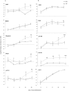
Figure 7. Time kinetics of MFRs and miRNA expression under VPA exposure.
mES cells were treated with 300 µM VPA from day 1 of neural differentiation. RNA samples were collected on day 5, 7, 9, 12, and 16 of differentiation. The expression of genes at each time point of differentiation is normalized to the expression in undifferentiated mES cells. The graphs demonstrate mean of log2 fold change in three independent biological replicates (differentiated vs. undifferentiated) ± SEM. (n = 3, t-test, *p<0.05, **p<0.01, ***p<0.001).
Figure 8
Figure 8. Expression of neuron- and myocyte-markers under VPA exposure.
Neural-differentiated ES cells were immuno-stained with neuron specific marker β-III-tubulin (red) and muscle specific marker α-actinin (green) after VPA (A) or PBS (B) treatment.
Figure 9
Figure 9. TSA induction of mir-206 and mir-10a during neural differentiation of mES cells.
The graph demonstrates mean of log2 fold change (TSA vs. solvent control) in two independent biological replicates ± SEM.

References

    1. Grandjean P, Landrigan PJ (2006) Developmental neurotoxicity of industrial chemicals. Lancet 368: 2167–2178 doi:10.1016/S0140-6736(06)69665-7 - DOI - PubMed
    1. Landrigan PJ (2010) What causes autism? Exploring the environmental contribution. Curr Opin Pediatr 22: 219–225 doi:10.1097/MOP.0b013e328336eb9a - DOI - PubMed
    1. Rice DD, Barone SS (2000) Critical periods of vulnerability for the developing nervous system: evidence from humans and animal models. Environ Health Perspect 108 Suppl 3 511–533. - PMC - PubMed
    1. Kuehn BM (2010) Increased Risk of ADHD Associated With Early Exposure to Pesticides, PCBs. JAMA 304: 27–28 doi:10.1001/jama.2010.860 - DOI - PubMed
    1. Sagiv SKS, Thurston SWS, Bellinger DCD, Tolbert PEP, Altshul LML, et al. (2010) Prenatal organochlorine exposure and behaviors associated with attention deficit hyperactivity disorder in school-aged children. Am J Epidemiol 171: 593–601 doi:10.1093/aje/kwp427 - DOI - PMC - PubMed

Publication types

MeSH terms