Review
. 2014 Jun 3;106(11):2291-304.
doi: 10.1016/j.bpj.2013.11.4498.

Vertex models of epithelial morphogenesis

Affiliations
Review

Vertex models of epithelial morphogenesis

Alexander G Fletcher et al. Biophys J. .

Abstract

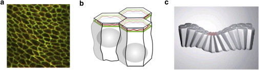
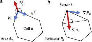
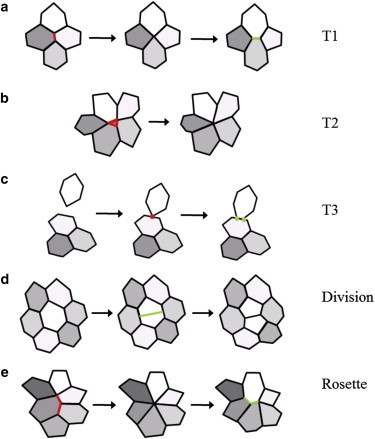
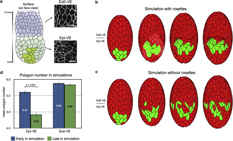
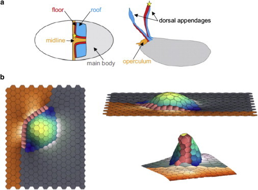
The dynamic behavior of epithelial cell sheets plays a central role during numerous developmental processes. Genetic and imaging studies of epithelial morphogenesis in a wide range of organisms have led to increasingly detailed mechanisms of cell sheet dynamics. Computational models offer a useful means by which to investigate and test these mechanisms, and have played a key role in the study of cell-cell interactions. A variety of modeling approaches can be used to simulate the balance of forces within an epithelial sheet. Vertex models are a class of such models that consider cells as individual objects, approximated by two-dimensional polygons representing cellular interfaces, in which each vertex moves in response to forces due to growth, interfacial tension, and pressure within each cell. Vertex models are used to study cellular processes within epithelia, including cell motility, adhesion, mitosis, and delamination. This review summarizes how vertex models have been used to provide insight into developmental processes and highlights current challenges in this area, including progressing these models from two to three dimensions and developing new tools for model validation.


Figures

Figure 1
Figure 1
Epithelial sheet morphology and organization. (a) Confocal microscope image of part of the follicular epithelium in Drosophila, with cell membranes visualized using an E-cadherin antibody, illustrating a typical polygonal cell packing geometry. (b) Schematic representation of the organization of neighboring cells in an epithelial sheet, connected via adhesion molecules (green) and cytoskeletal components (red) nearer their apical surface. (c) Schematic representation of epithelial bending associated with apical constriction. Images reproduced from Farhadifar et al. (24) and Lecuit et al. (2).
Figure 2
Figure 2
Illustration of key parameters in the explicit force- and energy-based models of vertex mechanics. (a) Forces acting on a given vertex i due to a given cell α associated with it, according to the explicit-force based model given by Eq. 2. (b) Forces acting on the vertex due to the cell according to Eq. 4, derived from the energy-based model given by Eq. 3.
Figure 3
Figure 3
Schematic diagram of junctional rearrangements, cell division, and cell removal in vertex models. (a) A T1 transition, in which two vertices sharing a short edge merge into a single vertex, which then decomposes into two new vertices such that the local network topology is changed. (b) A T2 transition, in which a cell shrinks to zero area and is removed, corresponding to delamination and/or apoptosis. (c) Example of a T3 transition, in a vertex/edge intersection is avoided by replacing the approaching vertex with two new vertices that are associated with the element. (d) Cell division, in which a parent cell is divided (in this case through its short axis) through the addition of two new vertices, resulting in two daughter cells. (e) Formation and resolution of multicellular rosettes, a generalization of T1 transitions involving a larger number of cells. (ae) Edges or vertices that are removed (red); edges or vertices that are created (green).
Figure 4
Figure 4
Simulation of AVE cell migration in the mouse VE with and without rosettes. (a) Schematic diagram of the visceral endoderm (VE) of a mouse egg-cylinder. The portions of the VE that cover the extraembryonic ectoderm (ExE-VE, blue) and that cover the epiblast (Epi-VE, green) as well as the proximally migrating AVE cells (dark green) are indicated. Differences in cell shapes are illustrated by high magnification views of the VE of an egg-cylinder stage mouse embryo stained with the tight junction marker ZO-1. (b) Snapshots of vertex model simulations of the mouse VE where rosettes are allowed to form (highlighted in gray), showing AVE cells (green) migrating in a single group. (c) As in panel b, but where rosettes are not allowed to form, showing AVE cells dispersing. (d) Comparison of mean polygon number in the ExE-VE and Epi-VE early and late in model simulations (corresponding to before and during AVE migration), recapitulating the experimentally observed reduction in mean polygon number in the Epi-VE. Images reproduced from Trichas et al. (23).
Figure 5
Figure 5
Simulation of three-dimensional epithelial morphogenesis in the developing Drosophila egg. (a) Schematic showing the locations of populations of different cell types within the follicular epithelium. (Left panel) Initial locations of the roof cells (blue), floor cells (red), midline cells (orange), and nonspecialized main body cells (gray). (Right panel) Final locations of these cell types relative to the completely formed eggshell. (b) (Clockwise from left) Top and side view of a vertex model simulation of out-of-plane tissue deformation, and final configuration showing a single completely formed appendage. Images reproduced from Osterfield et al. (63).

Similar articles

Cited by

References

    1. Martin A.C., Kaschube M., Wieschaus E.F. Pulsed contractions of an actin-myosin network drive apical constriction. Nature. 2009;457:495–499. - PMC - PubMed
    1. Lecuit T., Lenne P.F. Cell surface mechanics and the control of cell shape, tissue patterns and morphogenesis. Nat. Rev. Mol. Cell Biol. 2007;8:633–644. - PubMed
    1. Wallingford J.B. Neural tube closure and neural tube defects: studies in animal models reveal known knowns and known unknowns. Am. J. Med. Genet. C. Semin. Med. Genet. 2005;135C:59–68. - PubMed
    1. Lubarsky B., Krasnow M.A. Tube morphogenesis: making and shaping biological tubes. Cell. 2003;112:19–28. - PubMed
    1. Kiehart D.P., Galbraith C.G., Montague R.A. Multiple forces contribute to cell sheet morphogenesis for dorsal closure in Drosophila. J. Cell Biol. 2000;149:471–490. - PMC - PubMed

Publication types

LinkOut - more resources