

The NFL-TBS.40-63 anti-glioblastoma peptide disrupts microtubule and mitochondrial networks in the T98G glioma cell line
- PMID: 24896268
- PMCID: PMC4045719
- DOI: 10.1371/journal.pone.0098473
The NFL-TBS.40-63 anti-glioblastoma peptide disrupts microtubule and mitochondrial networks in the T98G glioma cell line
Abstract
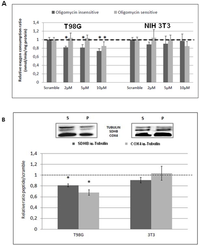
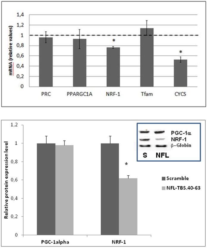
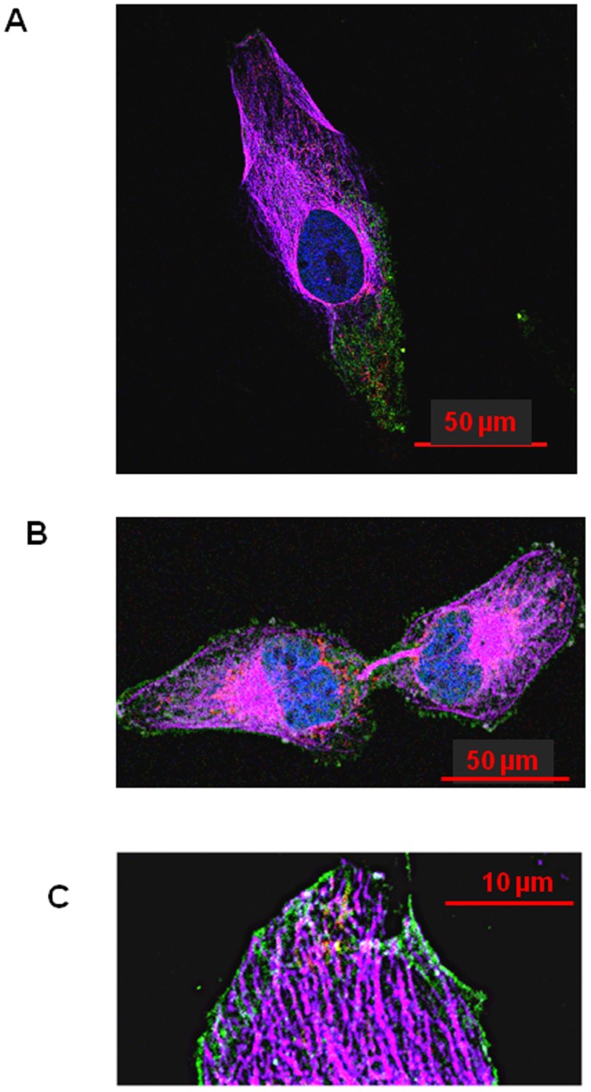
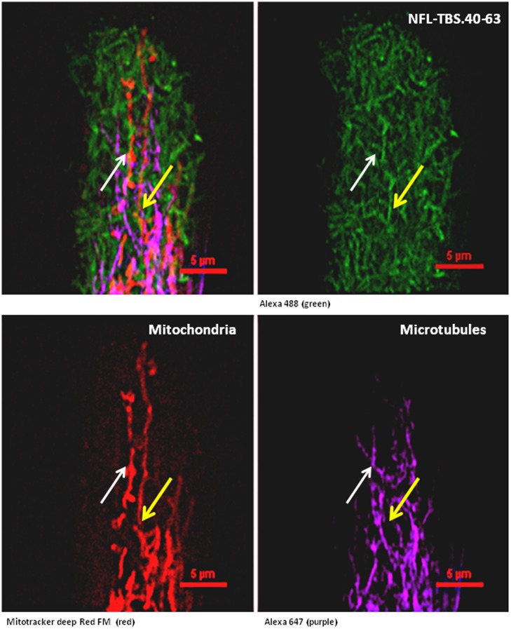
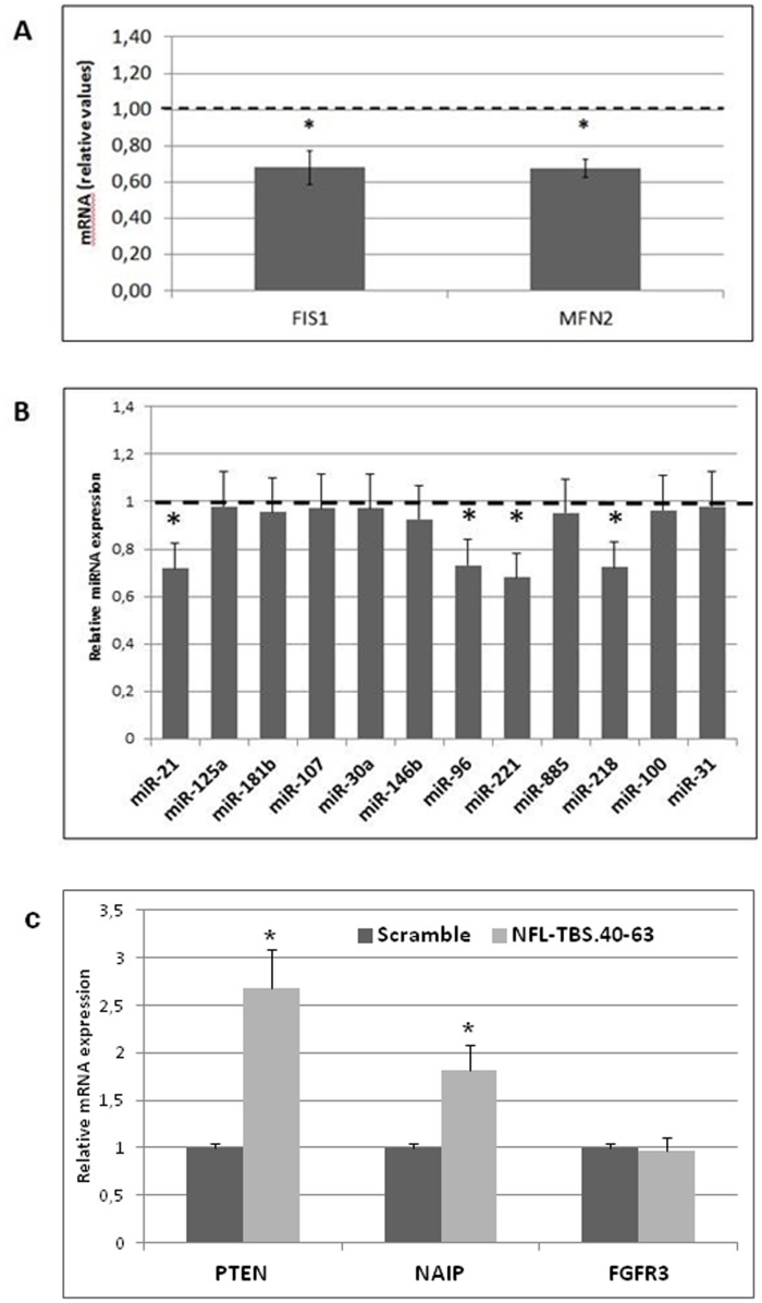
Despite aggressive therapies, including combinations of surgery, radiotherapy and chemotherapy, glioblastoma remains a highly aggressive brain cancer with the worst prognosis of any central nervous system disease. We have previously identified a neurofilament-derived cell-penetrating peptide, NFL-TBS.40-63, that specifically enters by endocytosis in glioblastoma cells, where it induces microtubule destruction and inhibits cell proliferation. Here, we explore the impact of NFL-TBS.40-63 peptide on the mitochondrial network and its functions by using global cell respiration, quantitative PCR analysis of the main actors directing mitochondrial biogenesis, western blot analysis of the oxidative phosphorylation (OXPHOS) subunits and confocal microscopy. We show that the internalized peptide disturbs mitochondrial and microtubule networks, interferes with mitochondrial dynamics and induces a rapid depletion of global cell respiration. This effect may be related to reduced expression of the NRF-1 transcription factor and of specific miRNAs, which may impact mitochondrial biogenesis, in regard to default mitochondrial mobility.
Conflict of interest statement
Figures
Similar articles
-
The NFL-TBS.40-63 anti-glioblastoma peptide enters selectively in glioma cells by endocytosis.Int J Pharm. 2013 Oct 1;454(2):738-47. doi: 10.1016/j.ijpharm.2013.04.004. Epub 2013 Apr 17. Int J Pharm. 2013. PMID: 23603097
-
The effect of functionalizing lipid nanocapsules with NFL-TBS.40-63 peptide on their uptake by glioblastoma cells.Biomaterials. 2013 Apr;34(13):3381-9. doi: 10.1016/j.biomaterials.2013.01.068. Epub 2013 Feb 4. Biomaterials. 2013. PMID: 23391494
-
A tubulin binding peptide targets glioma cells disrupting their microtubules, blocking migration, and inducing apoptosis.Mol Ther. 2012 Jul;20(7):1367-77. doi: 10.1038/mt.2012.45. Epub 2012 Apr 10. Mol Ther. 2012. PMID: 22491214 Free PMC article.
-
Emerging microtubule targets in glioma therapy.Semin Pediatr Neurol. 2015 Mar;22(1):49-72. doi: 10.1016/j.spen.2015.03.009. Epub 2015 Apr 4. Semin Pediatr Neurol. 2015. PMID: 25976261 Review.
-
Microtubule-targeted agents: when mitochondria become essential to chemotherapy.Biochim Biophys Acta. 2011 Jun;1807(6):679-88. doi: 10.1016/j.bbabio.2011.01.001. Epub 2011 Jan 7. Biochim Biophys Acta. 2011. PMID: 21216222 Review.
Cited by
-
Methods of miRNA delivery and possibilities of their application in neuro-oncology.Noncoding RNA Res. 2023 Oct 7;8(4):661-674. doi: 10.1016/j.ncrna.2023.10.002. eCollection 2023 Dec. Noncoding RNA Res. 2023. PMID: 37860265 Free PMC article. Review.
-
Biological activity of gold nanoparticles combined with the NFL-TBS.40-63 peptide, or with other cell penetrating peptides, on rat glioblastoma cells.Int J Pharm X. 2022 Sep 16;4:100129. doi: 10.1016/j.ijpx.2022.100129. eCollection 2022 Dec. Int J Pharm X. 2022. PMID: 36164551 Free PMC article.
-
Doxorubicin and NFL-TBS.40-63 peptide loaded gold nanoparticles as a multimodal therapy of glioblastoma.Discov Nano. 2025 Apr 28;20(1):72. doi: 10.1186/s11671-025-04249-z. Discov Nano. 2025. PMID: 40293574 Free PMC article.
-
NFL-TBS.40-63 Peptide Gold Complex Nanovector: A Novel Therapeutic Approach to Increase Anticancer Activity by Breakdown of Microtubules in Pancreatic Adenocarcinoma (PDAC).ACS Pharmacol Transl Sci. 2022 Oct 14;5(12):1267-1278. doi: 10.1021/acsptsci.2c00159. eCollection 2022 Dec 9. ACS Pharmacol Transl Sci. 2022. PMID: 36524008 Free PMC article.
-
Cell penetrating peptide (CPP) gold(iii) - complex - bioconjugates: from chemical design to interaction with cancer cells for nanomedicine applications.Nanoscale Adv. 2022 May 12;4(14):3010-3022. doi: 10.1039/d2na00096b. eCollection 2022 Jul 15. Nanoscale Adv. 2022. PMID: 36133522 Free PMC article.
References
-
- Stupp R, Mason WP, van den Bent MJ, Weller M, Fisher B, et al. (2005) Radiotherapy plus concomitant and adjuvant temozolomide for glioblastoma. N Engl J Med 352: 987–996. - PubMed
-
- Ohgaki H, Kleihues P (2005) Epidemiology and etiology of gliomas. Acta Neuropathol 109: 93–108. - PubMed
-
- Stupp R, Hegi ME, Mason WP, van den Bent MJ, Taphoorn MJ, et al. (2009) Effects of radiotherapy with concomitant and adjuvant temozolomide versus radiotherapy alone on survival in glioblastoma in a randomised phase III study: 5-year analysis of the EORTC-NCIC trial. Lancet Oncol 10: 459–466. - PubMed
Publication types
MeSH terms
Substances
LinkOut - more resources
Full Text Sources
Other Literature Sources
Medical
