. 2014 Jun 4;9(6):e98896.
doi: 10.1371/journal.pone.0098896. eCollection 2014.

Structure of the BTB domain of Keap1 and its interaction with the triterpenoid antagonist CDDO

Affiliations

Structure of the BTB domain of Keap1 and its interaction with the triterpenoid antagonist CDDO

Anne Cleasby et al. PLoS One. .

Abstract

The protein Keap1 is central to the regulation of the Nrf2-mediated cytoprotective response, and is increasingly recognized as an important target for therapeutic intervention in a range of diseases involving excessive oxidative stress and inflammation. The BTB domain of Keap1 plays key roles in sensing environmental electrophiles and in mediating interactions with the Cul3/Rbx1 E3 ubiquitin ligase system, and is believed to be the target for several small molecule covalent activators of the Nrf2 pathway. However, despite structural information being available for several BTB domains from related proteins, there have been no reported crystal structures of Keap1 BTB, and this has precluded a detailed understanding of its mechanism of action and interaction with antagonists. We report here the first structure of the BTB domain of Keap1, which is thought to contain the key cysteine residue responsible for interaction with electrophiles, as well as structures of the covalent complex with the antagonist CDDO/bardoxolone, and of the constitutively inactive C151W BTB mutant. In addition to providing the first structural confirmation of antagonist binding to Keap1 BTB, we also present biochemical evidence that adduction of Cys 151 by CDDO is capable of inhibiting the binding of Cul3 to Keap1, and discuss how this class of compound might exert Nrf2 activation through disruption of the BTB-Cul3 interface.


Conflict of interest statement

Competing Interests: The authors have read the journal's policy and have the following conflicts: The authors are currently employed by Astex Pharmaceuticals or GlaxoSmithKline. This does not alter the authors' adherence to PLOS ONE policies on sharing data and materials.

Figures

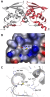
Figure 1
Figure 1. The structure of Keap1 BTB.
(A) Overall fold of the Keap1 BTB crystallographic dimer as a cartoon representation. The N and C-termini, and key alpha-helical secondary structural elements are labelled for one BTB monomer. The approximate position of Cys 151 is marked with an asterisk. (B) Surface around Cys 151 coloured according to electrostatic potential. Blue regions indicate areas of positive potential and red regions areas of negative potential as calculated by AstexViewer . (C) Details of Cys 151 environment showing close contact with Arg 135. Note that the side-chains of Lys 131 and and Lys 150 are highly flexible and exhibit very weak electron density. Some disorder is also evident for Arg 135.
Figure 2
Figure 2. CDDO and its interaction with the Keap1 domain of BTB.
(A) Chemical structure of CDDO/bardoxolone. (B) Surface representation of Keap1 BTB with CDDO bound in region of Cys 151. The final 2mFo-DFc electron density (contoured at 1σ) is shown as a green mesh. (C) Overview of CDDO binding in context of the BTB crystallographic dimer (only one binding site is shown for clarity). The surface of the CDDO binding site is shown in grey. (D) Details of covalent and non-covalent interactions between CDDO and BTB. Hydrogen bonds are denoted by dashed lines. The side-chain of Lys 131 is not visible in the electron density and has been truncated to the Cβ atom. (E) Schematic diagram of interactions between CDDO and BTB. His 129 is shown in the protonated state to emphasize its potential to form an electrostatic interaction with the carboxylate of CDDO. (F) Overlay of apo (white carbons) and CDDO-bound BTB (green carbons) in region of Cys 151.
Figure 3
Figure 3. Inhibitory effects of CDDO on Cul3 binding to wild-type C151 and C151S BTB-BACK as measured by AlphaScreen proximity assay.
The change in signal as a function of CDDO concentration is reported relative to a DMSO control, and is the mean of three measurements. Error bars represent the standard deviation.
Figure 4
Figure 4. Position of Keap1-bound CDDO in the context of possible Cul3 binding surfaces.
(A) Superposition of KLHL11/Cul3 (PDB code 4ap2) and BTB-CDDO showing proximity to Cul3 N-terminal tail (marked with arrow). KLHL11 is shown in a yellow cartoon representation, Cul3 in blue and the Keap1 BTB dimer in red and green. The CDDO binding site is highlighted as a grey surface, with the surface of CDDO shown in green. For clarity, bound CDDO is only shown for one BTB monomer, and a single copy of KLHL11/Cul3 is shown, although the KLHL11/Cul3 complex dimerizes through the KLHL11 BTB domain in the crystal lattice. (B) Surface representation of the KLHL11 Cul3 binding groove coloured by amino acid sequence identity with Keap1 (blue  =  identical, red  =  non-identical). Keap1 and KLHL11 sequences were aligned using ClustalW to determine regions of identity. The Cul3 N-terminal strand is depicted with blue carbons, and CDDO is shown as a surface representation in green. The position of CDDO was taken from that of its complex with BTB when overlaid with the KLHL11/Cul3 structure. The side-chain of Arg 19 is shown truncated to Cβ as deposited with the PDB. (C) Surface representation of BTB in the region of the CDDO binding groove, showing potential alternative path for Cul3 N-terminal tail upon binding to Keap1 (dashed arrow). The Cul3 and the IVR/BACK regions are taken from the superposition of the KLHL11/Cul3 structure with Keap1 BTB.
Figure 5
Figure 5. Comparison of Keap1 BTB C151W mutant with apo and CDDO-bound structures.
(A) Overlay of Keap1 BTB C151 (white carbons) and C151W mutant (red carbons) in Cys 151 region. The final 2mFo-DFc electron density (contoured at 1σ) is shown as a green mesh for the Trp 151 side-chain. (B) Overlay of the C151W BTB mutant (red carbons) and BTB-CDDO (green carbon) showing overlap between the antagonist and indole ring system of tryptophan. The volume occupied by the side-chain of Trp 151 has been highlighted as a white surface.

Similar articles

Cited by

References

    1. Zhang DD, Lo SC, Cross JV, Templeton DJ, Hannink M (2004) Keap1 is a redox-regulated substrate adaptor protein for a Cul3-dependent ubiquitin ligase complex. Mol Cell Biol 24: 10941–10953. - PMC - PubMed
    1. Itoh K, Chiba T, Takahashi S, Ishii T, Igarashi K, et al. (1997) An Nrf2/small Maf heterodimer mediates the induction of phase II detoxifying enzyme genes through antioxidant response elements. Biochem Biophys Res Commun 236: 313–322. - PubMed
    1. Wakabayashi N, Dinkova-Kostova AT, Holtzclaw WD, Kang MI, Kobayashi A, et al. (2004) Protection against electrophile and oxidant stress by induction of the phase 2 response: fate of cysteines of the Keap1 sensor modified by inducers. Proc Natl Acad Sci U S A 101: 2040–2045. - PMC - PubMed
    1. Zhang M, An C, Gao Y, Leak RK, Chen J, et al. (2013) Emerging roles of Nrf2 and phase II antioxidant enzymes in neuroprotection. Prog Neurobiol 100: 30–47. - PMC - PubMed
    1. Stogios PJ, Downs GS, Jauhal JJ, Nandra SK, Prive GG (2005) Sequence and structural analysis of BTB domain proteins. Genome Biol 6: R82. - PMC - PubMed

Publication types

MeSH terms

Substances