. 2014 Nov;39(12):2788-98.
doi: 10.1038/npp.2014.129. Epub 2014 Jun 4.

Control of the nigrostriatal dopamine neuron activity and motor function by the tail of the ventral tegmental area

Affiliations

Control of the nigrostriatal dopamine neuron activity and motor function by the tail of the ventral tegmental area

Romain Bourdy et al. Neuropsychopharmacology. 2014 Nov.

Abstract

Midbrain dopamine neurons are implicated in various psychiatric and neurological disorders. The GABAergic tail of the ventral tegmental area (tVTA), also named the rostromedial tegmental nucleus (RMTg), displays dense projections to the midbrain and exerts electrophysiological control over dopamine cells of the VTA. However, the influence of the tVTA on the nigrostriatal pathway, from the substantia nigra pars compacta (SNc) to the dorsal striatum, and on related functions remains to be addressed. The present study highlights the role played by the tVTA as a GABA brake for the nigrostriatal system, demonstrating a critical influence over motor functions. Using neuroanatomical approaches with tract tracing and electron microscopy, we reveal the presence of a tVTA-SNc-dorsal striatum pathway. Using in vivo electrophysiology, we prove that the tVTA is a major inhibitory control center for SNc dopamine cells. Using behavioral approaches, we demonstrate that the tVTA controls rotation behavior, motor coordination, and motor skill learning. The motor enhancements observed after ablation of the tVTA are in this regard comparable with the performance-enhancing properties of amphetamine, a drug used in doping. These findings demonstrate that the tVTA is a major GABA brake for nigral dopamine systems and nigrostriatal functions, and they raise important questions about how the tVTA is integrated within the basal ganglia circuitry. They also warrant further research on the tVTA's role in motor and dopamine-related pathological contexts such as Parkinson's disease.


Figures

Figure 1
Figure 1
The tVTA neurons project to the nigrostriatal system. (a) Injection of the anterograde tracer BDA into the tVTA and of the retrograde tracer CTb into the dorsolateral striatum (n=5). (b and c) tVTA terminals (arrows) contact dendrites and cell soma of striatum-projecting SNc neurons. (d) Injection of the anterograde tracer PhaL into the tVTA (n=4). (e and f) Electron micrographs showing tVTA axon terminals (tVTA-a) forming synapses (arrow) onto SNc dendrites labeled for tyrosine hydroxylase (TH-d) and contacted by additional unlabeled axons (ua) (f), larger view; (e), details of boxed area). (g) Quantitative analysis of the electron microscopy experiment. Most of the tVTA terminals synapse onto dendrites in the SNc that are immunoreactive for TH. BDA, biotinylated dextran amine; cc, corpus callosum; CTb, cholera toxin β-subunit; DLS, dorsolateral striatum; PhaL, Phaseolus vulgaris leucoagglutinin. Scale bars, 500 μm (a), 20 μm (b, c), 0.25 μm (e), 0.5 μm (f).
Figure 2
Figure 2
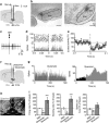
The tVTA controls SNc dopamine neuron activity. (a) Experimental protocol used for electrical stimulation. (b) Microphotographs of coronal sections through the SNc showing blue spots done at the end of recordings (left, anteromedial spot; right, posterolateral spot). (c) Example of a spike trace of a SNc dopamine neuron with a single pulse of tVTA stimulation at 1 mA. (d and e) Consequence of tVTA stimulation on a single SNc dopamine neuron (d) and on the mean of 19 neurons (e), n=19, F40,720=4.2, *p<0.05, ***p<0.001). (f) Experimental protocol used for chemical stimulation and inhibition of the tVTA. (g) Examples of unitary activities from single SNc dopamine neurons after tVTA chemical manipulation. (h) Injection site for muscimol-BODIPY in the tVTA. (i) Consequences of tVTA inhibition (muscimol, n=9) or excitation (glutamate, n=16) on SNc dopamine neuron activity (vs PBS vehicle, n=15) (Firing rate F2,37=22.3, p<0.001; bursting rate F2,37=23.6, p<0.001; spikes/burst F2,37=4.9, p<0.02; *p<0.05, ***p<0.001). Graphs represent mean±SEM. CLi, caudal linear nucleus of the raphe; IP, interpeduncular nucleus. Scale bar, 500 μm (b and h).
Figure 3
Figure 3
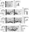
The tVTA controls rotation bias behavior. (a) Experimental protocol and histological evidence for unilateral tVTA lesion. (b) Unilateral tVTA lesion induces a contralateral rotation bias after amphetamine 3 mg/kg (control, n=17; lesion, n=12; **p<0.01). (c) Experimental protocol and histological evidence for 6-OHDA unilateral SNc lesion. (d) Unilateral SNc lesion induces an ipsilateral rotation bias after amphetamine administration (control, n=12; lesion, n=14; ***p<0.0001 referred to 50%). (e) Experimental protocol and histological evidence for unilateral VTA lesion with ibotenic acid. (f) Unilateral VTA lesion does not induce a rotation bias after amphetamine administration (control, n=9; lesion, n=7; referred to 50%). (g) Experimental protocol for unilateral SNc–tVTA lesions. The microphotograph illustrates a left side partial SNc lesion. (h) A tVTA lesion prevents the amphetamine-induced ipsilateral rotation bias observed after partial unilateral SNc lesion (SNc lesion, n=8; SNc–tVTA lesion, n=7; **p<0.01 referred to 50%). Graphs represent mean±SEM. 6-OHDA, 6-hydroxydopamine; IP, interpeduncular nucleus; PAG, periaqueductal gray; SNc, substantia nigra pars compacta; SNr, substantia nigra pars reticulata; TH, tyrosine hydroxylase; VTA, ventral tegmental area. Scale bars, 400 μm (a), 500 μm (c, e, g).
Figure 4
Figure 4
The tVTA controls motor behavior. (a) Experimental protocol and histological evidence for bilateral tVTA lesion. (b) Bilateral tVTA lesion increases SNc neuron basal firing (control, n=22; lesion, n=23; **p<0.01). (c) Locomotor activity of the bilateral tVTA-lesioned animals after subcutaneous saline injection (over 2 h) (control, n=9; lesion, n=10). (d) Number of falls in bilateral tVTA-lesioned and non-lesioned animals during their first rotarod training at low speed (5 r.p.m., 300 s) (control, n=9; lesion, n=10; ***p<0.001). (e) Motor performance of the bilateral tVTA-lesioned animals in a rotarod task at successively increasing fixed speeds (control, n=9; lesion, n=10; F3,51=5.1, p<0.01, ***p<0.001). (f) Overall performance comparison between bilateral tVTA-lesioned and non-lesioned animals, corresponding to e (**p<0.01). (g) Latency to fall and speed of bilateral tVTA-lesioned animals in a ramp rotarod task (0–45 r.p.m. in 120 s) (control, n=9; lesion, n=10; **p<0.01). (h) Motor skill learning of the bilateral tVTA-lesioned animals in a rotarod task measured over successive days (0–45 r.p.m., in 300 s) (control, n=10; lesion=9; F3,51=3.5, p<0.05; *p<0.05, **p<0.01). Graphs represent mean±SEM. r.p.m., rotations per minute; IP, interpeduncular nucleus. Scale bars, 500 μm (a).
Figure 5
Figure 5
Comparison of motor performances between tVTA and dorsolateral striatum lesions. (a) Experimental protocol and histological evidence for bilateral DLS lesion with 6-OHDA. (b) Latency to fall of bilateral tVTA and DLS-lesioned animals in a rotarod task at fixed speed (tVTA, n=10, DLS, n=11; referred to 100%, *p<0.05. (c) Number of falls during the first day of rotarod training at low speed (control, n=8–9; tVTA lesion, n=10; DLS lesion, n=11; **p<0.01 compared with controls; tVTA–DLS comparison, #p<0.001). (d) Overall performance comparison corresponding to Figure 5b (**p<0.01 referred to 100%). (e) Latency to fall in a ramp rotarod task (0–45 r.p.m. in 120 s) (tVTA, n=10; DLS, n=11; **p<0.01, #p=0.0507, referred to 100%). Graphs represent mean±SEM. ac, anterior commissure; cc, corpus callosum; DLS, dorsolateral striatum; r.p.m., rotations per minute; TH, tyrosine hydroxylase. Scale bar, 1 mm (a).
Figure 6
Figure 6
Amphetamine increases motor performance. (a) Amphetamine increases motor performance in a learned rotarod task (0–45 r.p.m., 120 s) (saline, n=10; amphetamine, n=10; *p<0.05). (b) Amphetamine increases motor skill learning in a rotarod task assessed over successive days (0–45 r.p.m., 180 s) (saline, n=12; amphetamine, n=13; F5,115=3.6, p<0.01, **p<0.01). Graphs represent mean±SEM.

Similar articles

Cited by

References

    1. Albin RL, Young AB, Penney JB. The functional anatomy of basal ganglia disorders. Trends Neurosci. 1989;12:366–375. - PubMed
    1. Akita H, Ogata M, Jitsuki S, Ogura T, Oh-Nishi A, Hoka S, et al. Nigral injection of antisense oligonucleotides to synaptotagmin I using HVJ-liposome vectors causes disruption of dopamine release in the striatum and impaired skill learning. Brain Res. 2006;1095:178–189. - PubMed
    1. Balcita-Pedicino JJ, Omelchenko N, Bell R, Sesack SR. The inhibitory influence of the lateral habenula on midbrain dopamine cells: ultrastructural evidence for indirect mediation via the rostromedial tegmental nucleus. J Comp Neurol. 2011;519:1143–1164. - PMC - PubMed
    1. Barrot M, Sesack SR, Georges F, Pistis M, Hong S, Jhou TC. Braking dopamine systems: a new GABA master structure for mesolimbic and nigrostriatal functions. J Neurosci. 2012;32:14094–14101. - PMC - PubMed
    1. Beeler JA, Cao ZF, Kheirbek MA, Ding Y, Koranda J, Murakami M, et al. Dopamine-dependent motor learning: insight into levodopa's long-duration response. Ann Neurol. 2010;67:639–647. - PMC - PubMed

Publication types

MeSH terms

Substances

LinkOut - more resources