

Abnormal brain iron metabolism in Irp2 deficient mice is associated with mild neurological and behavioral impairments
- PMID: 24896637
- PMCID: PMC4045679
- DOI: 10.1371/journal.pone.0098072
Abnormal brain iron metabolism in Irp2 deficient mice is associated with mild neurological and behavioral impairments
Abstract
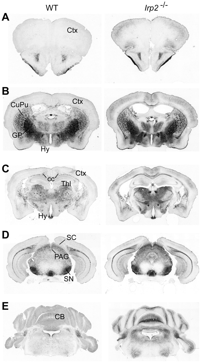
Iron Regulatory Protein 2 (Irp2, Ireb2) is a central regulator of cellular iron homeostasis in vertebrates. Two global knockout mouse models have been generated to explore the role of Irp2 in regulating iron metabolism. While both mouse models show that loss of Irp2 results in microcytic anemia and altered body iron distribution, discrepant results have drawn into question the role of Irp2 in regulating brain iron metabolism. One model shows that aged Irp2 deficient mice develop adult-onset progressive neurodegeneration that is associated with axonal degeneration and loss of Purkinje cells in the central nervous system. These mice show iron deposition in white matter tracts and oligodendrocyte soma throughout the brain. A contrasting model of global Irp2 deficiency shows no overt or pathological signs of neurodegeneration or brain iron accumulation, and display only mild motor coordination and balance deficits when challenged by specific tests. Explanations for conflicting findings in the severity of the clinical phenotype, brain iron accumulation and neuronal degeneration remain unclear. Here, we describe an additional mouse model of global Irp2 deficiency. Our aged Irp2-/- mice show marked iron deposition in white matter and in oligodendrocytes while iron content is significantly reduced in neurons. Ferritin and transferrin receptor 1 (TfR1, Tfrc), expression are increased and decreased, respectively, in the brain from Irp2-/- mice. These mice show impairments in locomotion, exploration, motor coordination/balance and nociception when assessed by neurological and behavioral tests, but lack overt signs of neurodegenerative disease. Ultrastructural studies of specific brain regions show no evidence of neurodegeneration. Our data suggest that Irp2 deficiency dysregulates brain iron metabolism causing cellular dysfunction that ultimately leads to mild neurological, behavioral and nociceptive impairments.
Conflict of interest statement
Figures


Similar articles
-
Severity of neurodegeneration correlates with compromise of iron metabolism in mice with iron regulatory protein deficiencies.Ann N Y Acad Sci. 2004 Mar;1012:65-83. doi: 10.1196/annals.1306.006. Ann N Y Acad Sci. 2004. PMID: 15105256
-
Tempol-mediated activation of latent iron regulatory protein activity prevents symptoms of neurodegenerative disease in IRP2 knockout mice.Proc Natl Acad Sci U S A. 2008 Aug 19;105(33):12028-33. doi: 10.1073/pnas.0805361105. Epub 2008 Aug 6. Proc Natl Acad Sci U S A. 2008. PMID: 18685102 Free PMC article.
-
Microcytic anemia, erythropoietic protoporphyria, and neurodegeneration in mice with targeted deletion of iron-regulatory protein 2.Blood. 2005 Aug 1;106(3):1084-91. doi: 10.1182/blood-2004-12-4703. Epub 2005 Apr 14. Blood. 2005. PMID: 15831703 Free PMC article.
-
Rare causes of hereditary iron overload.Semin Hematol. 2002 Oct;39(4):249-62. doi: 10.1053/shem.2002.35638. Semin Hematol. 2002. PMID: 12382200 Review.
-
Iron misregulation and neurodegenerative disease in mouse models that lack iron regulatory proteins.Neurobiol Dis. 2015 Sep;81:66-75. doi: 10.1016/j.nbd.2015.02.026. Epub 2015 Mar 11. Neurobiol Dis. 2015. PMID: 25771171 Free PMC article. Review.
Cited by
-
Iron Homeostasis in the CNS: An Overview of the Pathological Consequences of Iron Metabolism Disruption.Int J Mol Sci. 2022 Apr 19;23(9):4490. doi: 10.3390/ijms23094490. Int J Mol Sci. 2022. PMID: 35562883 Free PMC article. Review.
-
Candidate genes for COPD: current evidence and research.Int J Chron Obstruct Pulmon Dis. 2015 Oct 19;10:2249-55. doi: 10.2147/COPD.S80227. eCollection 2015. Int J Chron Obstruct Pulmon Dis. 2015. PMID: 26527870 Free PMC article. Review.
-
Biogenesis and functions of mammalian iron-sulfur proteins in the regulation of iron homeostasis and pivotal metabolic pathways.J Biol Chem. 2017 Aug 4;292(31):12744-12753. doi: 10.1074/jbc.R117.789537. Epub 2017 Jun 14. J Biol Chem. 2017. PMID: 28615439 Free PMC article. Review.
-
Mössbauer Spectra of Mouse Hearts Reveal Age-dependent Changes in Mitochondrial and Ferritin Iron Levels.J Biol Chem. 2017 Mar 31;292(13):5546-5554. doi: 10.1074/jbc.M117.777201. Epub 2017 Feb 15. J Biol Chem. 2017. PMID: 28202542 Free PMC article.
-
[Current insights into anemia in old age : Summary of the symposium "Anemia in old age" on the occasion of the annual congress of the German Society for Geriatrics (DGG) 2016 in Stuttgart].Z Gerontol Geriatr. 2018 Apr;51(3):343-348. doi: 10.1007/s00391-017-1212-8. Epub 2017 Apr 6. Z Gerontol Geriatr. 2018. PMID: 28386804 German.
References
Publication types
MeSH terms
Substances
Grants and funding
LinkOut - more resources
Full Text Sources
Other Literature Sources
Medical
Molecular Biology Databases
Miscellaneous
