Review
. 2014 May-Jun;3(3):193-210.
doi: 10.1002/wdev.135. Epub 2014 Apr 16.

Development of the embryonic and larval peripheral nervous system of Drosophila

Affiliations
Review

Development of the embryonic and larval peripheral nervous system of Drosophila

Aditi Singhania et al. Wiley Interdiscip Rev Dev Biol. 2014 May-Jun.

Abstract

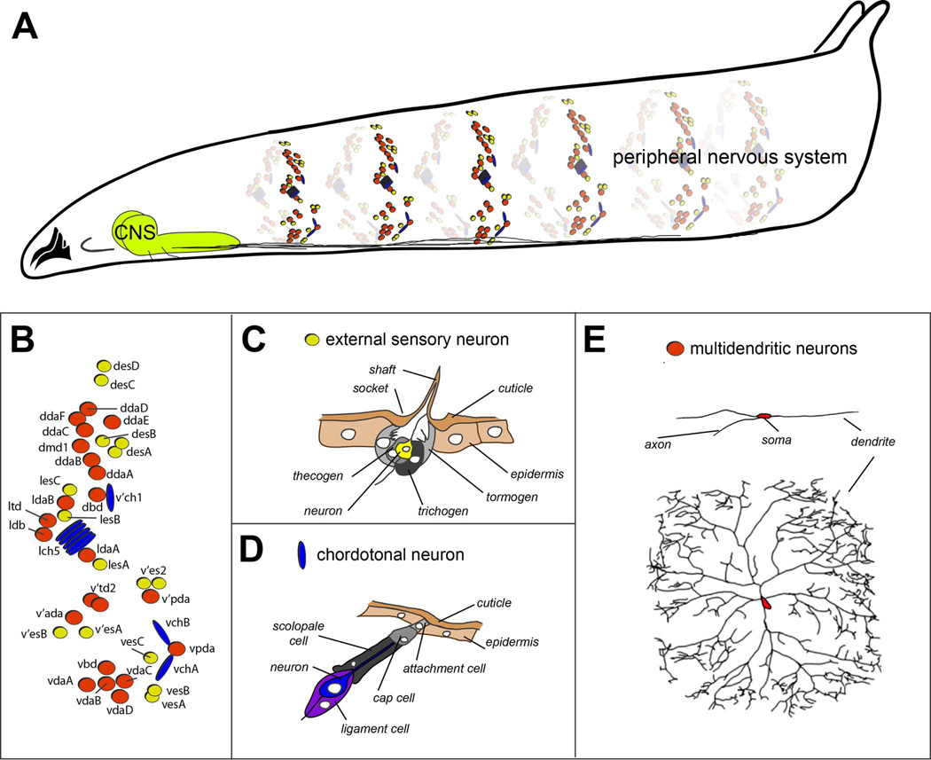
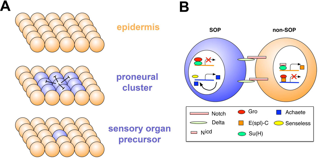
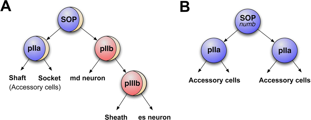
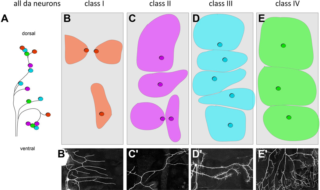
The peripheral nervous system (PNS) of embryonic and larval stage Drosophila consists of diverse types of sensory neurons positioned along the body wall. Sensory neurons, and associated end organs, show highly stereotyped locations and morphologies. Many powerful genetic tools for gene manipulation available in Drosophila make the PNS an advantageous system for elucidating basic principles of neural development. Studies of the Drosophila PNS have provided key insights into molecular mechanisms of cell fate specification, asymmetric cell division, and dendritic morphogenesis. A canonical lineage gives rise to sensory neurons and associated organs, and cells within this lineage are diversified through asymmetric cell divisions. Newly specified sensory neurons develop specific dendritic patterns, which are controlled by numerous factors including transcriptional regulators, interactions with neighboring neurons, and intracellular trafficking systems. In addition, sensory axons show modality specific terminations in the central nervous system, which are patterned by secreted ligands and their receptors expressed by sensory axons. Modality-specific axon projections are critical for coordinated larval behaviors. We review the molecular basis for PNS development and address some of the instances in which the mechanisms and molecules identified are conserved in vertebrate development.


Figures

Figure 1
Figure 1. Organization of the embryonic and larval peripheral nervous system of Drosophila
A. Drawing of a third instar Drosophila larva showing sensory elements that comprise the peripheral nervous system. For simplicity, only sensory neurons of a subset of abdominal segments are shown. Bundled sensory axons project to the central nervous system (CNS) that resides in the ventral and anterior part of the larva. B. Schematic of the arrangement of sensory neurons in a single abdominal hemisegment. External sensory organs are indicated by yellow circles, chordotonal organs by blue ovals, and multidendritic neurons by red circles. C. Drawing of external sensory organ structure. Names of individual cellular elements are indicated. Drawing adapted, with permission, from Comprehensive Molecular Insect Science. Vol. 1: Hartenstein V. Development of Insect Sensilla. pp. 379–419, 2005. D. Drawing of chordotonal organ structure. Names of individual cellular elements are indicated. Drawing adapted, with permission, from Comprehensive Molecular Insect Science. Vol. 1: Hartenstein V. Development of Insect Sensilla. pp. 379–419, 2005. E. Tracings of multidendritic neurons. Two different neurons are shown, the dorsal bipolar dendrite neuron (top), and a class IV nociceptive neuron (bottom). Note the different degrees of dendritic branching shown by the two neurons. Tracing of class IV neuron reproduced, with permission from Grueber et al., 2003.
Figure 2
Figure 2. Sensory organ precursor specification
A. Proneural clusters (blue spheres) typified by expression of basic helix-loop-helix transcription factors, arise from a field of equipotential epidermal cells (orange). From the proneural cluster a single sensory organ precursor emerges, which inhibits proneural gene expression in surrounding cells, a process termed lateral inhibition. B. Molecular basis for lateral inhibition. Delta ligand and Notch receptor are expressed in future SOP and non-SOP cells. Delta levels become higher in the SOP, which promotes higher Notch signaling in non-SOP cells. The Notch intracellular domain collaborates with the Suppressor of Hairless protein to promote expression of Enhancer of split in non-SOP cells. Enhancer of split acts together with the Groucho transcriptional repressor to shut off expression of the proneural gene achaete. In the future SOP, Groucho and Suppressor of hairless repress expression of Enhancer of split, so that proneural gene expression can persist. High-level expression of proneural genes is promoted by Senseless and also by proneural factors themselves in a positive feedback loop. Drawing adapted with permission from (Castro et al. Development, 2005).
Figure 3
Figure 3. Asymmetric cell division in the SOP lineage diversifies cell fates
A. Wildtype external sensory organ precursor lineage (ESOP). Tan arc in the SOP, pIIb, and pIIIb represents a crescent of asymmetrically localized Numb protein. The SOP divides to produce pIIa (blue) and pIIb (red) cells. pIIb inherits Numb and undergoes another round of numb-dependent asymmetric cell division which gives rise to an md neuron and pIIIb (red). pIIIb in turn generates a sheath cell and external sensory (es) neuron. The pIIa cell generates accessory cells, the shaft cell and socket cell. B. In the absence of numb the SOP divides into two identical pIIa cells, which divide to give rise to accessory cells. No neurons are produced by SOPs mutant for numb.
Figure 4
Figure 4. Distinct dendritic morphologies and territories of different classes of dendritic arborization (da) neurons
A. Schematic of arrangement of dendritic arborization neuron cell bodies. Different morphological classes of neurons are color coded red = class I, purple = class II, light blue = class III, and green = class IV. B-E. Schematics of the territories occupied by the different classes of da neurons in each hemisegment. The approximate locations of sensory neuron cell bodies within each class are indicated by circles. The approximate dendritic coverage of the neuron is indicated by colored areas. Note tiling among class III neurons and among class IV neurons. B'-E' Representative dendritic arbors of class I (B'), class II (C'), class III (D') and class IV (E') neurons. Neuron in C' adapted with permission from (Matthews et al., 2007).
Figure 5
Figure 5. Molecular mechanisms that specify the location of dendritic branches along an arbor
A, B. Images of class IV ddaC neurons that are wildtype (A) and mutant for dlic2 (B). The field size of dlic2 mutant neurons and dendritic arbors are abnormal (yellow arrowheads). The dlic2 mutant neurons show a shift in dendritic branches from distal to proximal (indicated by dotted blue squares). The red arrowheads indicate sensory axons, which are also thickened in dlic2 mutants. Reproduced with permission from Zheng et al., 2008. C. Schematic representing Golgi outposts (red) and Rab5 early endosomes (Rab5-EE, blue). D. Close up view of a dendritic branch indicating dynein-dependent microtubule minus (−) end directed movement of organelles to distal dendritic branches.
Figure 6
Figure 6. Self-avoidance control by Dscam1
A. Schematic drawing of sensory dendrites in wild-type and Dscam1 loss of function mutants. Overlaps between sister branches are indicated by red arrows. B. The Dscam1 locus consists of a series of exon cassettes (exon 4, 6, 9, and 17) that via alternative splicing can together generate over 38,000 distinct isoforms. Exons 4, 6, and 9 code for subregions of extracellular Ig domains 2 (red) and 3 (blue), or the entire Ig7 (green), respectively. Exon 17 generates either of two alternative transmembrane domains (yellow). Numbers in parentheses indicate the number of potential forms of each alternatively spliced domain. Flattened blue ovals indicate FN3 domains. C. In wild-type larvae overlap is observed between dendritic branches of two different dendritic arborization sensory neurons, schematized as green and magenta neurons. Note the extensive crossing between branches of different neurons (arrows), but no overlap between branches of the same cell. Overexpression of a single Dscam1 isoform in cells that normally overlap leads to repulsion and separation of their fields such that few crossings among branches are observed.
Figure 7
Figure 7. Tiling of da neuron dendritic arbors
Tiling between adjacent dendrites leads to non-overlapping dendritic fields and complete territory coverage. Shown is a confocal micrograph of class IV da neurons in a second instar larva. The cell bodies, axons, and dendrites of four different neurons are painted different colors to illustrate the tiling between them.
Figure 8
Figure 8. Sensory axon targeting in the CNS
A. Organization and specification of axonal projections of md and ch organ sensory neurons. The ventral nerve cord is organized as an outer cell body region and an inner neuropil comprised of sensory axons, motor dendrites and interneuron processes. Axon tracts labeled by Fasciclin II (a cell adhesion molecule) are distributed along the dorsoventral and mediolateral axes and provide landmarks for description of axon targeting. The axons of some multidendritic neurons (blue) target a medial region of the neuropil. Chordotonal neurons (red) target intermediate neuropil. Mutations in robo3 shift chordotonal projections to a medial position that is characteristic of multidendritic neurons. l, lateral; i, intermediate; m, medial. Drawings based on data from Zlatic et al., 2003 . B. Semaphorin (Sema)/Plexin (Plex) signaling in the assembly of sensory circuitry. Top graph diagram shows relative levels of Sema 2a (green) and Sema 2b (yellow) across the mediolateral axis of the neuorpil. High levels of Sema-2a are seen at the midline, and lower levels laterally. Sema-2a mediates repulsion of PlexB-expressing axons from the midline region. Sema-2b is found at high levels in medial and intermediate axon tracts. Lower drawing shows projection pattern of Sema-2b-expressing interneuron to medial and intermediate tracts. This interneuron source of Sema-2b attracts PlexB-expressing chordotonal organs to the intermediate fascicle. Drawings adapted with permission from Wu et al., 2011 .

Similar articles

Cited by

References

    1. McIver SB. Mechanoreception. In: Kerkut GA, Gilbert LI, editors. Comprehensive Insect Physiology Biochemistry and Pharmacology. Vol. 6. Oxford: Elsevier; 1985. pp. 71–132.
    1. Hartenstein V. Development of Insect Sensilla. In: Gilbert LI, Iatrou K, Gill SS, editors. Comprehensive Molecular Insect Science. Vol. 1. Elsevier; 2005. pp. 379–419.
    1. Bodmer R, Jan YN. Morphological differentiation of the embryonic peripheral neurons in Drosophila. Roux's Arch Dev Biol. 1987;196:69–77. - PubMed
    1. Grueber WB, Jan LY, Jan YN. Tiling of the Drosophila epidermis by multidendritic sensory neurons. Development. 2002;129:2867–2878. - PubMed
    1. Sweeney NT, Li W, Gao FB. Genetic manipulation of single neurons in vivo reveals specific roles of flamingo in neuronal morphogenesis. Dev Biol. 2002;247:76–88. - PubMed

Publication types

MeSH terms

LinkOut - more resources