. 2014 Sep 15;462(3):555-65.
doi: 10.1042/BJ20140076.

Selenoprotein S is involved in maintenance and transport of multiprotein complexes

Affiliations

Selenoprotein S is involved in maintenance and transport of multiprotein complexes

Anton A Turanov et al. Biochem J. .

Abstract

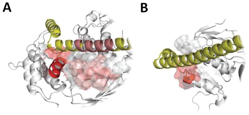
SelS (Selenoprotein S) is a selenocysteine-containing protein with roles in ER (endoplasmic reticulum) function and inflammation. It has been implicated in ERAD (ER-associated protein degradation), and clinical studies revealed an association of its promoter polymorphism with cytokine levels and human diseases. However, the pathways and interacting proteins that could shed light on pathogenesis of SelS-associated diseases have not been studied systematically. We performed a large-scale affinity isolation of human SelS and its mutant forms and analysed the proteins that interact with them. All previously known SelS targets and nearly two hundred additional proteins were identified that were remarkably enriched for various multiprotein complexes. Subsequent chemical cross-linking experiments identified the specific interacting sites in SelS and its several targets. Most of these interactions involved coiled-coil domains. The data suggest that SelS participates in intracellular membrane transport and maintenance of protein complexes by anchoring them to the ER membrane.


Figures

Figure 1
Figure 1. Design of the study to identify SelS targets
A) Schematic representations of SelS fusion proteins. Various forms of HHT-tagged SelS were cloned into pCI-ToxoSECIS vector resulting in HHT-SelS CU, HHT-SelS CC and HHT-SelS C constructs. SelS constructs without the coiled-coil domain (HHT-SelS-ΔCC) or containing only the cytosolic part of SelS without H2-Kb (HT-SelS-CT) were prepared as described in the Experimental section. An asterisk shows the site for Eco47III restriction enzyme introduced in the SelS sequence. B) Schematic representation of the SelS target search. HeLa cells were transfected [1] with various SelS constructs, subjected to fluorescent microscopy analysis [2.1]. HEK 293T cells were transfected [1] with the same constructs and used for large-scale affinity purification of SelS and its binding proteins on anti-HA-agarose followed by elution with TEV protease [2.2, 3, 4]. The eluted proteins were subjected to SDS-PAGE [5], and prepared [6,7] for LC-MS/MS [7] or cross-linking [8]. Mass-spectrometry data were filtered and analyzed [9] by Sequest and CompPASS to generate the SelS target list used for cross-linking analysis by xQuest. Crosslinked proteins were verified by Western blotting [10].
Figure 2
Figure 2. Analysis of SelS-binding proteins
A) Silver staining of SDS-PAGE performed on SelS samples eluted with TEV protease (corresponding to step 5 in Figure 1B). HEK 293T cells were left untransfected (lane 1) or transfected with the constructs coding for HHT-SelS CU (lane 2), HHT-SelS-ΔCC (lane 3) and HT-SelS-CT (lane 4). B) SDS-PAGE analysis of SelS target preparations. SelS samples and control (SelS CU, SelS C, HEK untransfected) were fractionated by SDS-PAGE, and proteins were stained with silver staining. Positions of TEV protease and SelS are indicated by arrows on right and molecular weights in kDa are shown on left. C) Flow chart illustrating analysis of mass-spec data for identification of SelS-interacting proteins. D) Western blotting of SelS samples shown in Figure 2A with the corresponding antibodies (SelS-top panel, p97, SelK and Derlin 1- bottom panels). Positions of SelS forms are indicated by arrows on right and molecular weights in kDa are shown on left.
Figure 3
Figure 3. Cellular localization of SelS
A) HeLa cells were transfected with the construct coding for the ER-targeted H2-Kb-HA-TEV-SelS. B) HeLa cells were transfected with the construct coding for the ER-targeted H2Kb-HA-TEV-SelS that lacked the coiled-coil domain. C) HeLa cells were transfected with the construct coding for the ER-targeted H2K-HA-TEV-SelS that lacked the transmembrane domain (TM). D) HeLa cells were transfected with the construct coding for the ER-targeted H2K-HA-TEV-SelS. E) HeLa cells were transfected with the construct coding for the ER-targeted H2K-HA-TEV-SelS that lacked the coiled-coil domain. Transfected cells were stained with SelS or HA antibodies (green) and KDEL antibodies (red) (left and middle panels). Nuclei were stained with DAPI (blue) and shown in the merged images (right panels).
Figure 4
Figure 4. Isolation of SelS-interacting protein complexes
A) HEK 293T cells were transfected with pCI-ToxoSECIS (vector) or with various HA-tagged SelS forms (see descriptions in the text). The SelS interacting proteins were isolated on HA-agarose and specific binding of SelS targets was assessed by Western blotting under non-reducing conditions with SelS (upper panel), SelK (middle panel) or p97 (lower panel) antibodies. Position of SelS dimers is shown with asterisk. B) Known proteins involved in ERAD were visualized by STRING and the identified SelS targets indicated by red circles. Proteins with cross-links to SelS are highlighted with a black circle. C) Previously identified SelS targets were visualized by STRING, and the SelS targets found in the current study are highlighted with red circles. D) SelS interacts with diverse multiprotein complexes. Protein complexes were visualized by STRING. SelS targets are indicated by red circles. Proteins with cross-links to SelS are highlighted with black circles. OST – oligosaccharyltransferase complex, MS – multisynthetase complex, APC – anaphase-promoting complex, NP – nuclear pore complex.
Figure 5
Figure 5. Analysis of SelS-interacting proteins identified by cross-linking
A) HEK 293 cells were transfected with HA-tagged SelS (HHT-SelS CC), and SelS targets isolated on HA-agarose and treated with a cross-linker. Efficiency of cross-linking was analyzed with SelS (left panel) and p97 (middle panel) antibodies. Coomassie Brilliant Blue (CBB) stained membrane is shown on right. Molecular weight markers in kDa are shown on left. B–E) SelS samples (initial - input, flow-through – flow, final – IP:HA) prior to crosslinking were analyzed by Western blotting with (B) EPRS, (C) AIMP2, (D) USP5 and (E) NCKAP1 antibodies. Actin served as a control. F) Coomassie Blue stained membrane (upper panel) and Western blotting with SelS antibodies (lower panel) are shown on right. G) SelS was immunoprecipitated and the samples analyzed by Western blotting with indicated antibodies. Arrows (on right of each blot) indicate the expected migration of proteins (indicated on left of each blot). Input – initial sample. IP:control – sample treated as IP:SelS except that no SelS antibodies were added during the procedure. Flow - sample after treatment with SelS antibodies. IP:SelS – proteins immunoprecipitated with SelS antibodies.
Figure 6
Figure 6. Binding modes of SelS
Two representative proteins have been selected among SelS targets for docking calculations: one containing a coiled coil domain (p97 ATPase, PDB structure: 3CF3; panel A) and one without it (CYB5R3/NADH-cytochrome b5 reductase 3, PDB structure: 1UMK; panel B). In both panels, SelS is shown in yellow, and the docking partner (p97, CYB5R3) in light grey (secondary structure only shown, for clarity). Contact regions of binding partners are presented as molecular surfaces; linked regions (from cross-link experiments, SRVAILKANL in p97, and RNKHSA in CYB5R3) are shown in bright red. Finally (panel A only), dark red colored sections of both SelS and p97 highlight interactions directly involving coiled coil domains in both proteins.

References

    1. Hatfield DL, Gladyshev VN. How selenium has altered our understanding of the genetic code. Mol Cell Biol. 2002;22:3565–3576. - PMC - PubMed
    1. Kryukov GV, Castellano S, Novoselov SV, Lobanov AV, Zehtab O, Guigo R, Gladyshev VN. Characterization of mammalian selenoproteomes. Science. 2003;300:1439–1443. - PubMed
    1. McCann JCA, Bruce N. Adaptive dysfunction of selenoproteins from the perspective of the triage theory: why modest selenium deficiency may increase risk of diseases of aging. FASEB Journal. 2011;25:1793–1814. - PubMed
    1. Papp LV, Holmgren A, Khanna KK. Selenium and selenoproteins in health and disease. Antioxid Redox Signal. 2010;12:793–795. - PubMed
    1. Ye Y, Shibata Y, Yun C, Ron D, Rapoport TA. A membrane protein complex mediates retro-translocation from the ER lumen into the cytosol. Nature. 2004;429:841–847. - PubMed

Publication types

MeSH terms