. 2014 Jun 4;9(6):e98655.
doi: 10.1371/journal.pone.0098655. eCollection 2014.

Comprehensive secondary structure elucidation of four genera of the family Pospiviroidae

Affiliations

Comprehensive secondary structure elucidation of four genera of the family Pospiviroidae

Tamara Giguère et al. PLoS One. .

Abstract

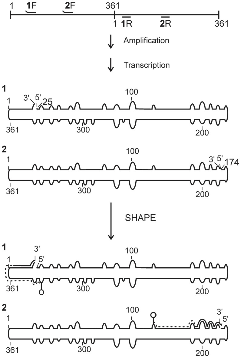
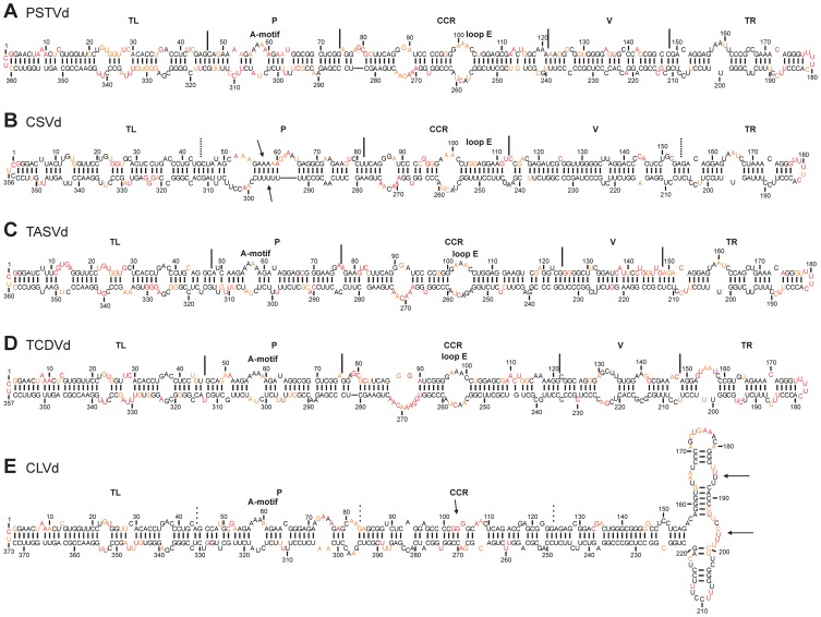
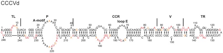
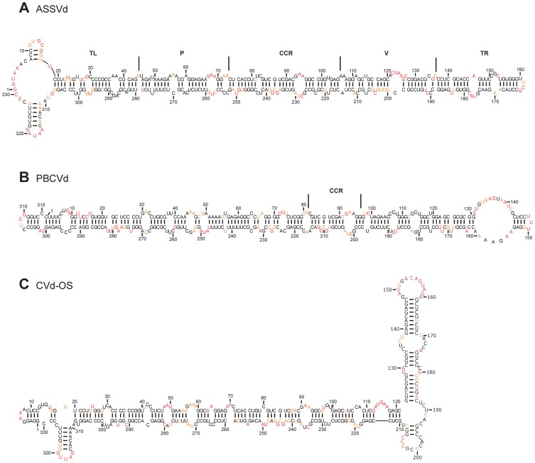
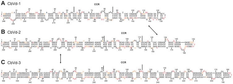
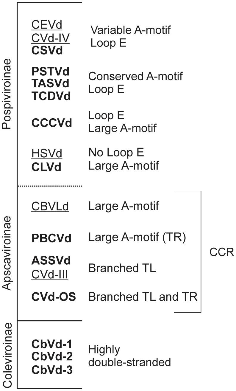
Viroids are small, circular, single stranded RNA molecules that infect plants. Since they are non-coding, their structures play a critical role in their life cycles. To date, little effort has been spend on elucidating viroid structures in solution due to both the experimental difficulties and the time-consuming nature of the methodologies implicated. Recently, the technique of high-throughput selective 2'-hydroxyl acylation analyzed by primer extension (SHAPE) was adapted for the probing of the members of family Avsunviroidae, all of whom replicate in the chloroplast and demonstrate ribozyme activity. In the present work, twelve viroid species belonging to four different genera of the family Pospiviroidae, whose members are characterized by the presence of a central conserved region (CCR) and who replicate in nucleus of the host, were probed. Given that the structures of five distinct viroid species from the family Pospiviroidae have been previously reported, an overview of the different structural characteristics for all genera and the beginning of a manual classification of the different viroids based on their structural features are presented here.


Conflict of interest statement

Competing Interests: The authors have declared that no competing interests exist.

Figures

Figure 1
Figure 1. Schematic flow chart illustrating the detailed steps of the hSHAPE experiment with dimeric PSTVd.
The numbered primers are used for the amplification of the PSTVd monomers (i.e. 1F/1R and 2F/2R). The raised ends of the primers represent the non-complementary regions, specifically the T7 RNA polymerase promoter, which is used for the run-off transcription. The RNAs obtained after amplification and transcription are numbered 1 and 2. These RNAs were subjected to the SHAPE reaction followed by primer extension. During the latter step the reverse transcriptase produces a cDNA fragment (dashed line) the length of the distance from the start of the RNA until the first adduct (e.g. circle on top of a line).
Figure 2
Figure 2. The most stable structures for the viroids from the genus Pospiviroid.
The final structural models of PSTVd (A), CSVd (B), TASVd (C), TCVd (D) and, CLVd (E) obtained by SHAPE were folded with the RNAstructure software. The nucleotides in black denote those of low SHAPE reactivities (0–0.40). Those in orange are of intermediate reactivities (0.40–0.85) and those in red are highly reactive (>0.85). The different regions are either delimited (full lines), or approximated (dashed lines), and the presence of both the A-motif and the loop E are noted.
Figure 3
Figure 3. The most stable structure for a viroid from the genus Cocadviroid.
The final structural model of CCCVd obtained by SHAPE and folded by the RNAstructure software. The nucleotides in black denote those of low SHAPE reactivities (0–0.40) Those in orange are of intermediate reactivities (0.40–0.85) and those in red are highly reactive (>0.85). The different regions are delimited by the full lines.
Figure 4
Figure 4. The most stable structures for the viroids from the genus Apscaviroid.
The final structural models for ASSVd (A), PBCVd (B) and CVd-OS (C) obtained by SHAPE and folded by the RNAstructure software. The nucleotides in black denote those of low SHAPE reactivities (0–0.40). Those in orange are of intermediate reactivities (0.40–0.85) and those in red are highly reactive (>0.85). The CCR are delimited by the full lines.
Figure 5
Figure 5. The most stable structures for the viroids from the genus Coleviroid.
The final structural models for CbVd-1 (A), CbVd-2 (B) and CbVd-3 (C) obtained by SHAPE and folded by the RNAstructure software. The nucleotides in black denote those of low SHAPE reactivities (0–0.40). Those in orange are of intermediate reactivities (0.40–0.85) and those in red are highly reactive (>0.85). The CCR are delimited by the full lines. The arrows indicate the sequences that are identical in the three viroids.
Figure 6
Figure 6. Index of the viroids with SHAPE elucidated structures, classified according to their characteristics.
The viroids are also classified by their subfamilies. Viroids in bold are those reported in this article. The underlined names represent viroids whose structures were elucidated in other reports . The characteristics described in the paper are listed on the right.

References

    1. Ding B (2010) Viroids: Self-replicating, mobile, and fast-evolving noncoding regulatory RNAs. Wiley Interdiscip Rev RNA 1: 362–375. - PubMed
    1. Diener TO (2003) Discovering viroids—a personal perspective. Nat Rev Microbiol 1: 75–80. - PubMed
    1. Flores R, Hernandez C, Martinez de Alba AE, Daros JA, Di Serio F (2005) Viroids and viroid-host interactions. Annu Rev Phytopathol 43: 117–139. - PubMed
    1. Bussiere F, Lafontaine D, Perreault JP (1996) Compilation and analysis of viroid and viroid-like RNA sequences. Nucleic Acids Res 24: 1793–1798. - PMC - PubMed
    1. Haseloff J, Symons RH (1981) Chrysanthemum stunt viroid: Primary sequence and secondary structure. Nucleic Acids Res 9: 2741–2752. - PMC - PubMed

Publication types

LinkOut - more resources