. 2014 Jun 4;9(6):e98946.
doi: 10.1371/journal.pone.0098946. eCollection 2014.

Catalytic activity of cGMP-dependent protein kinase type I in intact cells is independent of N-terminal autophosphorylation

Affiliations

Catalytic activity of cGMP-dependent protein kinase type I in intact cells is independent of N-terminal autophosphorylation

Raghavan Vallur et al. PLoS One. .

Abstract

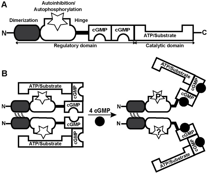
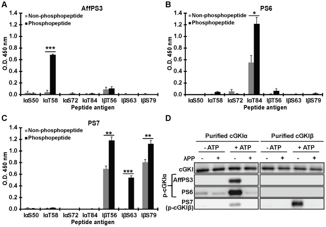
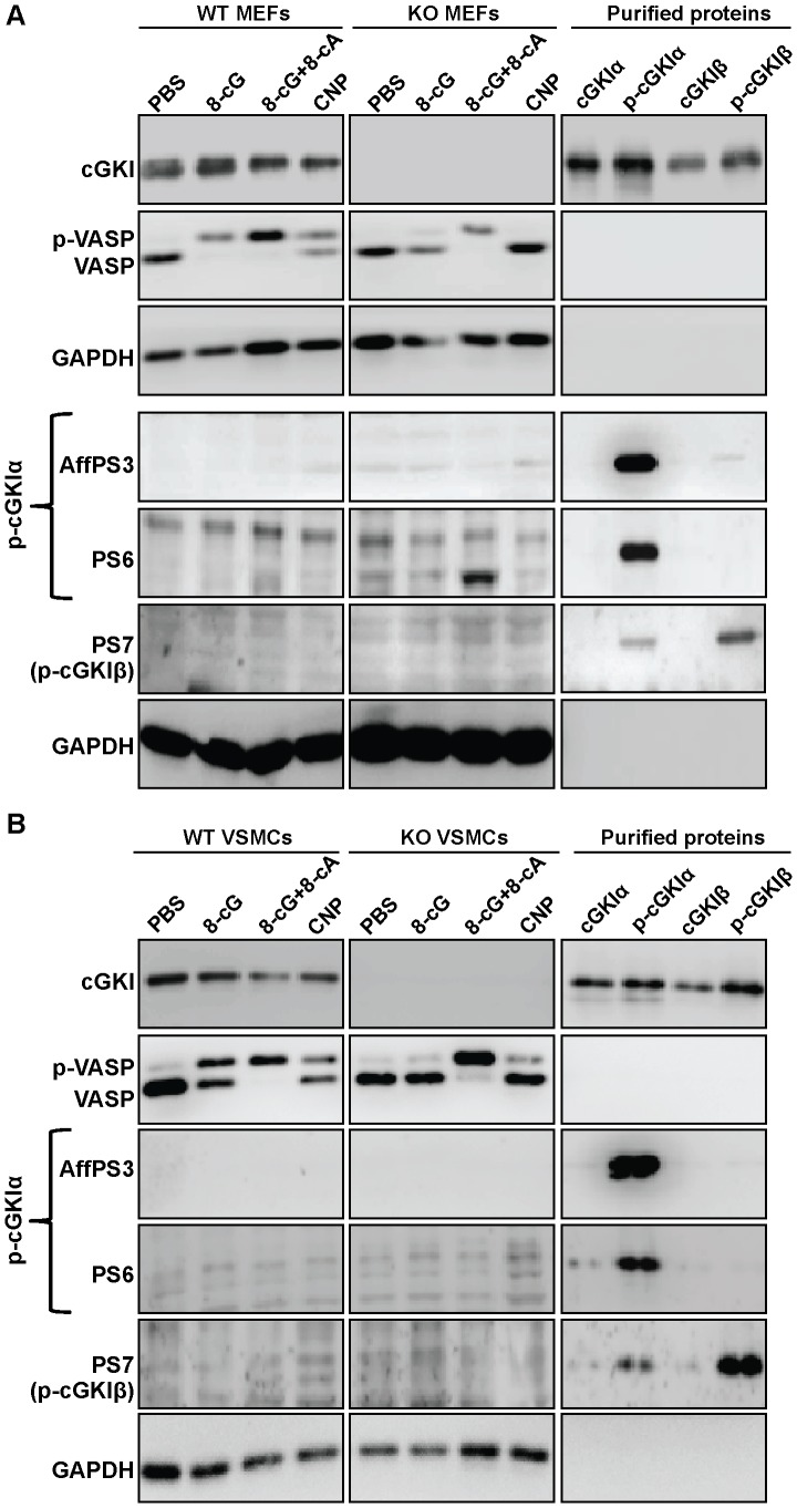
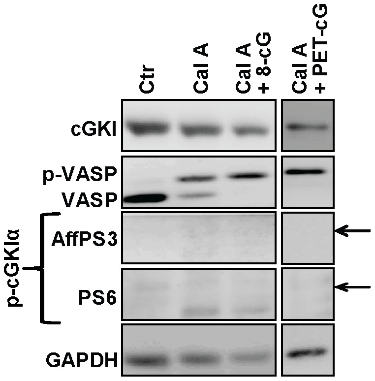
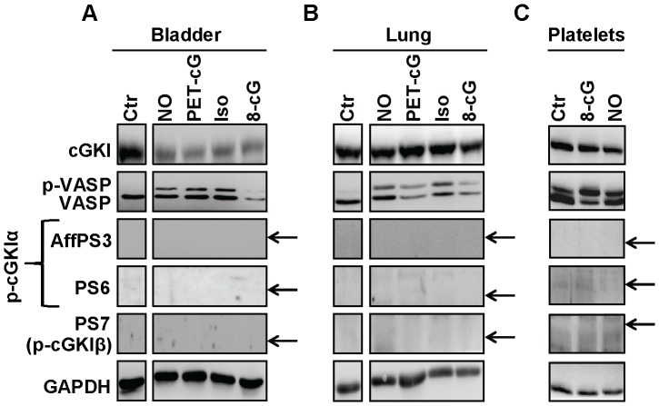
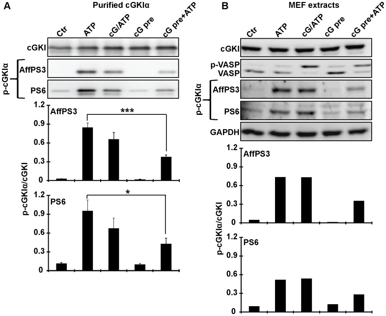
Although cGMP-dependent protein kinase type I (cGKI) is an important mediator of cGMP signaling and upcoming drug target, its in vivo-biochemistry is not well understood. Many studies showed that purified cGKI autophosphorylates multiple sites at its N-terminus. Autophosphorylation might be involved in kinase activation, but it is unclear whether this happens also in intact cells. To study cGKI autophosphorylation in vitro and in vivo, we have generated phospho-specific antisera against major in vitro-autophosphorylation sites of the cGKI isoforms, cGKIα and cGKIβ. These antisera detected specifically and with high sensitivity phospho-cGKIα (Thr58), phospho-cGKIα (Thr84), or phospho-cGKIβ (Thr56/Ser63/Ser79). Using these antisera, we show that ATP-induced autophosphorylation of cGKI in purified preparations and cell extracts did neither require nor induce an enzyme conformation capable of substrate heterophosphorylation; it was even inhibited by pre-incubation with cGMP. Interestingly, phospho-cGKI species were not detectable in intact murine cells and tissues, both under basal conditions and after induction of cGKI catalytic activity. We conclude that N-terminal phosphorylation, although readily induced in vitro, is not required for the catalytic activity of cGKIα and cGKIβ in vivo. These results will also inform screening strategies to identify novel cGKI modulators.


Conflict of interest statement

Competing Interests: The authors have declared that no competing interests exist.

Figures

Figure 1
Figure 1. General structure and current working model of cGKI.
(A) cGKI consists of a C-terminal catalytic domain and an N-terminal regulatory domain. The catalytic domain contains binding sites for ATP and protein substrates with Ser/Thr residues. The regulatory domain comprises two non-identical cGMP-binding pockets and additional regions with multiple functions: a leucine zipper for dimerization of two identical subunits, an overlapping autoinhibitory/autophosphorylation region (open star), and a flexible hinge region connecting the N-terminal region to the rest of the protein. (B) According to the current model, the homodimeric enzyme cannot heterophosphorylate substrates in the absence of cGMP (left). Binding of cGMP (black circles) results in a conformational change that allows heterophosphorylation of substrates (right). According to in vitro studies with purified cGKI, the N-terminal region of the inactive kinase is not phosphorylated (left, stars), and activation is associated with autophosphorylation of distinct sites in this region (right, star with a “P”). However, it is not clear whether or not N-terminal phosphorylation of cGKI does also occur in intact cells (right, star with a “?”).
Figure 2
Figure 2. Validation of phospho-specific antisera by ELISAs with antigenic peptides (A–C) and Western blotting with purified proteins (D).
Three polyclonal rabbit antisera were analyzed for their specificity and sensitivity to detect distinct phospho-sites of cGKIα (affinity-purified antiserum AffPS3, and non-purified antiserum PS6) or cGKIβ (non-purified antiserum PS7). (A–C) ELISAs were used to test binding of the antisera to non-phosphorylated (grey bars) and phosphorylated (black bars) peptide antigens (for peptide IDs and sequences, see Table 1). Data shown are means from three independent experiments ± SEM; *p≤0.05, **p≤0.01, ***p≤0.001. (D) Western blot detection of autophosphorylated cGKIα and cGKIβ by the antisera. Purified cGKI isoforms were incubated in the absence or presence of 0.1 mM ATP for 15 min at 30°C. Aliquots of the reactions were subsequently treated with lambda protein phosphatase (λPP) for 90 min at 30°C. Proteins (20 ng) were separated on SDS gels and Western blots were probed with a pan-(nonphospho-specific) cGKI antibody that detects both cGKIα and cGKIβ in their non-phosphorylated state (upper panels), and with the newly generated phospho-specific antisera. Data shown in D are representative for at least three independent experiments.
Figure 3
Figure 3. Analysis of N-terminal cGKI phosphorylation in intact MEFs (A) and VSMCs (B).
Cells from wild-type (WT) and cGKI-knockout (KO) mice were incubated for 15 min at 37°C under control conditions (PBS), or in the presence of 1 mM 8-Br-cGMP (8-cG), 1 mM 8-Br-cGMP and 10 mM 8-Br-cAMP (8-cG+8-cA), or 100 nM CNP. Then, cells were lysed in denaturating buffer and cell lysates were subjected to Western blot analysis with a pan-(nonphospho-specific) cGKI antibody, anti-VASP, anti-GAPDH, and the phospho-specific antisera AffPS3, PS6, and PS7. Phosphorylation of VASP at Ser157 (p-VASP) was monitored by immunodetection of the band shift to a higher apparent molecular weight. The protein amounts of MEF lysates loaded were 15 µg for immunostaining with anti-cGKI and anti-VASP, and 50 µg for immunodetection with the phospho-specific antisera; 12 µg of VSMC lysates were loaded; GAPDH was used as respective loading control. Purified proteins (4 ng) were loaded as controls for non-phosphorylated (cGKIα, cGKIβ) and autophosphorylated (p-cGKIα, p-cGKIβ) cGKI isoforms. Similar results were obtained in three independent experiments.
Figure 4
Figure 4. Effect of inhibition of protein Ser/Thr phosphatases on N-terminal cGKI phosphorylation in intact cells.
Wild-type MEFs were incubated at 37°C under control conditions (1% DMSO in PBS for 15 min; Ctr), or for 15 min in the presence of 100 nM of the PP1/PP2A inhibitor, calyculin A (Cal A), or for 15 min in the presence of 100 nM calyculin A followed by 15 min with 1 mM 8-Br-cGMP (Cal A+8-cG) or 1 mM 8-Br-PET-cGMP (Cal A+PET-cG). Then the cells were lysed in denaturating buffer and cell lysates (10 µg) were analyzed by Western blotting with the indicated antibodies. GAPDH was used as loading control. The arrows indicate the positions expected for phospho-cGKI species as determined by co-loading of purified proteins on the same gel. Similar results were obtained in three independent experiments.
Figure 5
Figure 5. Analysis of N-terminal cGKI phosphorylation in native mouse tissues and platelets.
(A) Bladder and (B) lung were rapidly isolated from wild-type mice and then incubated in Tyrode buffer for 15 min at room temperature under control conditions (Ctr) or in the presence of 100 nM calyculin A and 0.1 mM DEA-NONOate (NO), 1 mM 8-Br-PET-cGMP (PET-cG), 0.01 mM isoprenaline hydrochloride (Iso), or 1 mM 8-Br-cGMP (8-cG). (C) Platelets were isolated from wild-type mice and incubated for 10 min at 37°C under control conditions (Ctr) or in the presence of 1 mM 8-Br-cGMP (8-cG) or 3 mM DEA-NONOate (NO). Lysates (22 µg for bladder, 30 µg for lung, and equal fractions by volume for platelets) were subjected to Western blot analysis with the indicated antibodies. GAPDH was used as loading control. The arrows indicate the positions expected for phospho-cGKI species as determined by co-loading of purified proteins on the same gel. The displayed results are representative for three independent experiments.
Figure 6
Figure 6. N-terminal phosphorylation of cGKI in purified preparations (A) and cell extracts (B).
(A) Purified cGKIα or (B) cell extracts prepared from wild-type MEFs in non-denaturating buffer were incubated for 15 min at 30°C under control conditions (Ctr) or in the presence of 0.1 mM ATP or 0.1 mM ATP combined with 0.1 mM cGMP (cG/ATP). Alternatively, samples were pre-incubated for 15 min at 30°C with 0.1 mM cGMP. Then they were further incubated either under control conditions without ATP (cG pre) or in the presence of 0.1 mM ATP (cG pre+ATP) for another 15 min at 30°C. Purified proteins (20 ng) or cell extracts (10 µg) were analyzed for N-terminal phosphorylation of cGKIα by Western blotting with antisera AffPS3 and PS6. The total amount of cGKI was detected with a pan-(nonphospho-specific) cGKI antibody, and phospho-VASP in cell extracts was monitored with anti-VASP antibody. GAPDH was used as loading control for cell extracts. Below the Western blots, the semiquantitative densitometric analysis of phospho-cGKI signals is shown. It was performed using ImageJ software and is given as the ratio of the intensity of the phospho-band detected by AffPS3 or PS6 (p-cGKI) divided by the intensity of the respective cGKI band detected by the pan-cGKI antibody in the same sample (cGKI). Data shown in the bar graphs in (A) are means ± SEM (n = 3 independent experiments); *p≤0.05, ***p≤0.001. Data shown in the bar graphs in (B) are means of 2 independent experiments.

Similar articles

Cited by

References

    1. Beavo JA, Brunton LL (2002) Cyclic nucleotide research — still expanding after half a century. Nat Rev Mol Cell Biol 3: 710–718. - PubMed
    1. Kemp-Harper B, Feil R (2008) Meeting report: cGMP matters. Sci Signal 1: pe12. - PubMed
    1. Feil R, Lohmann SM, de Jonge H, Walter U, Hofmann F (2003) Cyclic GMP-dependent protein kinases and the cardiovascular system: insights from genetically modified mice. Circ Res 93: 907–916. - PubMed
    1. Hofmann F, Feil R, Kleppisch T, Schlossmann J (2006) Function of cGMP-dependent protein kinases as revealed by gene deletion. Physiol Rev 86: 1–23. - PubMed
    1. Francis SH, Busch JL, Corbin JD (2010) cGMP-dependent protein kinases and cGMP phosphodiesterases in nitric oxide and cGMP action. Pharmacol Rev 62: 525–563. - PMC - PubMed

Publication types

MeSH terms

LinkOut - more resources