. 2014 Jun 4:15:69.
doi: 10.1186/1471-2202-15-69.

Defined α-synuclein prion-like molecular assemblies spreading in cell culture

Affiliations

Defined α-synuclein prion-like molecular assemblies spreading in cell culture

Suzana Aulić et al. BMC Neurosci. .

Abstract

Background: α-Synuclein (α-syn) plays a central role in the pathogenesis of synucleinopathies, a group of neurodegenerative disorders that includes Parkinson disease, dementia with Lewy bodies and multiple system atrophy. Several findings from cell culture and mouse experiments suggest intercellular α-syn transfer.

Results: Through a methodology used to obtain synthetic mammalian prions, we tested whether recombinant human α-syn amyloids can promote prion-like accumulation in neuronal cell lines in vitro. A single exposure to amyloid fibrils of human α-syn was sufficient to induce aggregation of endogenous α-syn in human neuroblastoma SH-SY5Y cells. Remarkably, endogenous wild-type α-syn was sufficient for the formation of these aggregates, and overexpression of the protein was not required.

Conclusions: Our results provide compelling evidence that endogenous α-syn can accumulate in cell culture after a single exposure to exogenous α-syn short amyloid fibrils. Importantly, using α-syn short amyloid fibrils as seed, endogenous α-syn aggregates and accumulates over several passages in cell culture, providing an excellent tool for potential therapeutic screening of pathogenic α-syn aggregates.


Figures

Figure 1
Figure 1
In vitro conversion and AFM characterization of three different recombinant α-syn assemblies. Kinetics for the formation of β-sheet-rich assemblies: human α-syn oligomer (dotted black line), human α-syn short fibrils (red triangle line) and human α-syn long fibrils (gray dotted line), and control (gray solid line). (A) Red arrows indicate the collection time of the aggregates. (B) The lag phase, in hours, of all β-sheet structure preparations was measured using Thioflavin T assay. The lag phase distribution of α-syn and FLAG-α-syn amyloid preparations showed no difference (P > 0.5), indicating that the presence of FLAG-tag did not affect fibril formation. (C) AFM imaging analysis was performed at the end of the fibrillization reactions. AFM scan topographical images of α-syn deposited on mica surface. (D) The size of particles was measured: the typical height of α-syn oligomers was 0.65 ± 0.11 nm, the height of short and log fibrils was 2.79 ± 1.20 nm, and 6.08 ± 1.38 nm, respectively. The values are the average calculated on 20 fibrils. The error is the standard deviation.
Figure 2
Figure 2
Cytotoxicity, extracellular α-syn uptake and recruitment of endogenous α-syn in SH-SY5Y cells. (A) Cells were treated with 5 μg/mL α-syn oligomers, short, and long fibrils for 24 hours. Results are mean ± st. dev. of three independent experiments performed in six replicas. (B) Internalization of FLAG-α-syn amyloids into neuroblastoma cells SH-SY5Y. α-Syn deposition (green) was detected by anti-human α-syn antibody. Immunofluorescence was performed on cells exposed to FLAG-α-syn amyloids (oligomers, short amyloid fibrils and long fibrils) for 7 days. Bar, 11.9 μm. (C) Formation of endogenous α-syn deposits induced by extracellular α-syn short amyloid fibrils. Cells were treated for 7 days. Western blot analysis was performed using the α-syn (C-20)-R antibody, which recognizes endogenous human α-syn in SH-SY5Y cells. The nuclei (blue) were stained with DAPI. Scale bars, 11.9 μm.
Figure 3
Figure 3
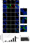
Infection of the non-transfected SH-SY5Y cells with α-syn short amyloid fibrils and sub-passing over time. (A) Cells were infected with recombinant human FLAG-α-syn short fibrils. Cells were cultured on coverslips for each passage (p0 to p6). The deposition of exogenous short amyloid fibrils FLAG-α-syn (red) was detected by anti-FLAG antibody. Human endogenous α-syn detected by anti-human α-syn (C-20)-R antibody (green). The nuclei were stained with DAPI (blue). Bar, 12 μm. On the right p4, p5, and p6 zoomed images. Corrected total cell fluorescence (CTCF) from immunofluorescence imaging shows the induction of endogenous α-syn in SH (neuroblastoma SH-SY5Y cell line) infected with human α-syn short amyloid fibrils during the passages (bottom panel). The analysis was performed on at least 150 cells, n = 3, ***p < 0.005. Values are mean ± SD. (B) PCR analyses directed to α-syn using the total RNA extracted from non-transfected SH-SY5Y cells. Mix, RT-, S. Mix: negative controls, pET11A: positive control, SH-SF p6: SH-SY5Y cells passaged six times after exposure to short amyloid fibrils, SH: SH-SY5Y cells not treated with short amyloid fibrils. α-Syn was detected after 25 cycles and did not show any significant difference in transcripts before and after infection with short amyloid fibrils of recombinant α-syn.
Figure 4
Figure 4
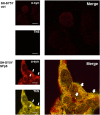
Thioflavin-S (ThS) positive staining of endogenous α-syn aggregates. SH-SY5Y cells were infected with recombinant human α-syn short amyloid fibrils (SF: short amyloid fibrils of α-syn) and sub-passed at sixth passage (p6). The deposition and level of α-syn (red) in α-syn-infected cell lines after six passages were detected by anti-α-syn antibody (Additional file 6: Table S2). The colocalization was observed between the ThS signal (yellow) and anti-human α-syn antibody (red). The nuclei (blue) were stained with DAPI. Scale bars, 12 μm.
Figure 5
Figure 5
Aggregation of α-syn in dopaminergic human cell lines infected with recombinant human α-syn short amyloid fibrils and sub-passaged over five passages (p5). (A) Western blot of different assemblies formed in vitro. (B) Western blot of cell lysates (SH: SH-SY5Y cells; SH-OE SH-SY5Y overexpressing human α-syn); Ctrl: non-treated cells. SFp5: α-syn short amyloid fibril-infected cells at fifth passage. HMW: High molecular weight aggregates. (C) Western blot analysis of aggregated SH-OE cell lysates treated with α-syn short amyloid fibrils, and non-treated (Ctrl); Total: total cell lysates. Sequential extraction of α-syn aggregates in 1% Triton X-100 lysis buffer (S-TX), followed by 2% SDS lysis buffer (S). (P): a pellet fraction after the pellet was treated with 2% SDS. Anti-human α-syn (C-20)-R antibody was used (Additional file 6: Table S2).
Figure 6
Figure 6
Short amyloid fibrils induce the formation of phosphorylated α-syn aggregates over passages. (A) Presence of phosphorylated α-syn aggregates in SH-SY5Y overexpressing α-syn (SH-OE, top left panel). From left to right: neuroblastoma SH-SY5Y (SH), SH-SY5Y infected with human α-syn short amylod fibrils at passage 9 (SH-SFp9), SH-SY5Y overexpressing α-syn (SH-OE) and SH-SY5Y overexpressing α-syn-infected with human α-syn short amylod fibrils at passage 9 (SH-OE-SFp9). Cells were immunolabeled with anti-α-syn antibody (green, upper panel) or anti-phospho S129 α-syn Ab59264 (green, lower panel). Scale bars, 5 μm. Arrows point at α-syn aggregates. Images are representative of at least three independent experiments. (B) High percentage of phosphorylated α-syn aggregates in SH-SY5Y α-syn-infected cells. Bar diagram showing quantification of α-syn aggregates (left) and phospho S129 α-syn aggregates (right) in SH-SY5Y α-syn cells and SH-SY5Y α-syn-infected cells. ***p < 0.001 (Student t test, n > 150 cells counted for each condition per experiment). Error bars represent the SEM.

References

    1. Spillantini MG, Schmidt ML, Lee VM, Trojanowski JQ, Jakes R, Goedert M. Alpha-synuclein in Lewy bodies. Nature. 1997;15(6645):839–840. doi: 10.1038/42166. - DOI - PubMed
    1. Wakabayashi K, Yoshimoto M, Tsuji S, Takahashi H. Alpha-synuclein immunoreactivity in glial cytoplasmic inclusions in multiple system atrophy. Neurosci Lett. 1998;15(2–3):180–182. - PubMed
    1. Spillantini MG, Crowther RA, Jakes R, Cairns NJ, Lantos PL, Goedert M. Filamentous alpha-synuclein inclusions link multiple system atrophy with Parkinson’s disease and dementia with Lewy bodies. Neurosci Lett. 1998;15(3):205–208. doi: 10.1016/S0304-3940(98)00504-7. - DOI - PubMed
    1. Tu PH, Galvin JE, Baba M, Giasson B, Tomita T, Leight S, Nakajo S, Iwatsubo T, Trojanowski JQ, Lee VM. Glial cytoplasmic inclusions in white matter oligodendrocytes of multiple system atrophy brains contain insoluble alpha-synuclein. Ann Neurol. 1998;15(3):415–422. doi: 10.1002/ana.410440324. - DOI - PubMed
    1. Uversky VN, Li J, Fink AL. Evidence for a partially folded intermediate in alpha-synuclein fibril formation. J Biol Chem. 2001;15(14):10737–10744. doi: 10.1074/jbc.M010907200. - DOI - PubMed

Publication types